Dana Farber Cancer Institute, Belfer Institute of Cancer Science, Boston, MA, USA.
Department of Respiratory Medicine and Clinical Immunology, Graduate School of medicine, Osaka University, Osaka, Japan.
J Immunother Cancer. 2019 Feb 6;7(1):32. doi: 10.1186/s40425-019-0504-5.
Tumor orchestrated metabolic changes in the microenvironment limit generation of anti-tumor immune responses. Availability of arginine, a semi-essential amino acid, is critical for lymphocyte proliferation and function. Levels of arginine are regulated by the enzymes arginase 1,2 and nitric oxide synthase (NOS). However, the role of arginase activity in lung tumor maintenance has not been investigated in clinically relevant orthotopic tumor models.
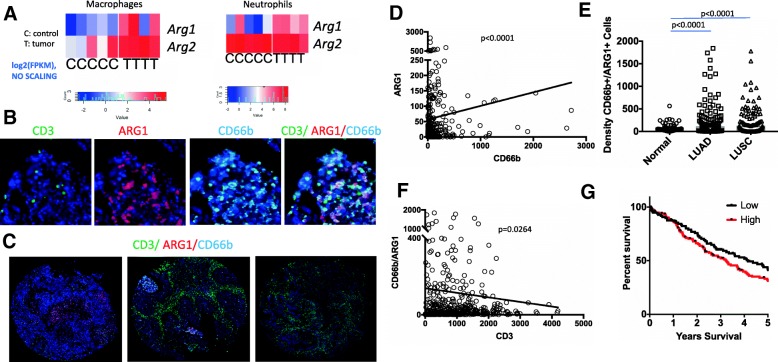
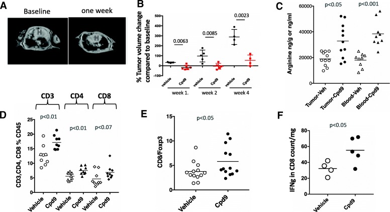
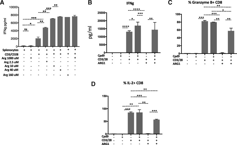
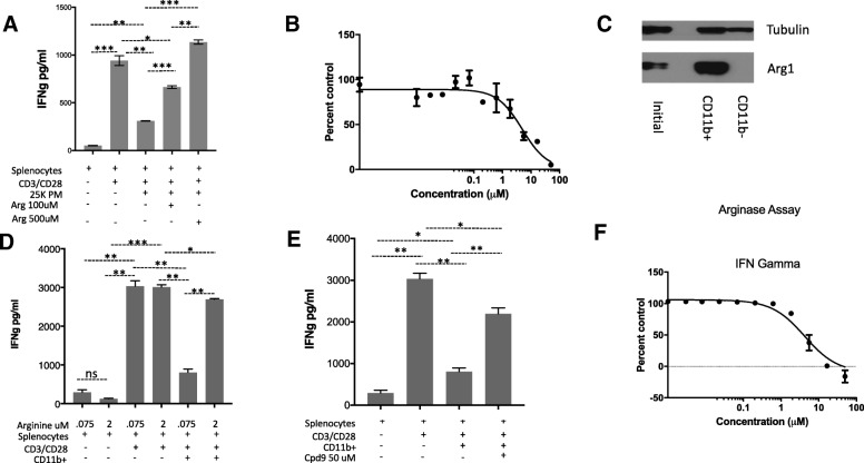
RNA sequencing (RNA-seq) of sorted cell populations from mouse lung adenocarcinomas derived from immunocompetent genetically engineered mouse models (GEMM)s was performed. To complement mouse studies, a patient tissue microarray consisting of 150 lung adenocarcinomas, 103 squamous tumors, and 54 matched normal tissue were stained for arginase, CD3, and CD66b by multiplex immunohistochemistry. Efficacy of a novel arginase inhibitor compound 9 in reversing arginase mediated T cell suppression was determined in splenocyte ex vivo assays. Additionally, the anti-tumor activity of this compound was determined in vitro and in an autochthonous immunocompetent Kras GEMM of lung adenocarcinoma model.
Analysis of RNA-seq of sorted myeloid cells suggested that arginase expression is elevated in myeloid cells in the tumor as compared to the normal lung tissue. Accordingly, in the patient samples arginase 1 expression was mainly localized in the granulocytic myeloid cells and significantly elevated in both lung adenocarcinoma and squamous tumors as compared to the controls. Our ex vivo analysis demonstrated that myeloid derived suppressor cell (MDSC)s cause T cell suppression by arginine depletion, and suppression of arginase activity by a novel ARG1/2 inhibitor, compound 9, led to restoration of T cell function by increasing arginine. Treatment of Kras GEMM of lung cancer model with compound 9 led to a significant tumor regression associated with increased T cell numbers and function, while it had no activity across several murine and human non-small cell (NSCLC) lung cancer lines in vitro.
We show that arginase expression is elevated in mouse and patient lung tumors. In a KRAS GEMM arginase inhibition diminished growth of established tumors. Our data suggest arginase as an immunomodulatory target that should further be investigated in lung tumors with high arginase activity.
肿瘤在微环境中引发的代谢变化限制了抗肿瘤免疫反应的产生。精氨酸是一种半必需氨基酸,其可用性对淋巴细胞的增殖和功能至关重要。精氨酸的水平受精氨酸酶 1、2 和一氧化氮合酶 (NOS) 的调节。然而,在临床上相关的原位肿瘤模型中,尚未研究肺肿瘤维持过程中精氨酸酶活性的作用。
对源自免疫活性遗传工程小鼠模型 (GEMM) 的小鼠肺腺癌分离细胞群体进行 RNA 测序 (RNA-seq)。为了补充小鼠研究,对由 150 例肺腺癌、103 例鳞状肿瘤和 54 例匹配的正常组织组成的患者组织微阵列进行了精氨酸酶、CD3 和 CD66b 的多重免疫组织化学染色。在脾细胞体外实验中,测定新型精氨酸酶抑制剂化合物 9 逆转精氨酸酶介导的 T 细胞抑制的效果。此外,还在体外和肺腺癌的自发免疫活性 Kras GEMM 模型中测定了该化合物的抗肿瘤活性。
对分离的髓样细胞 RNA-seq 的分析表明,与正常肺组织相比,肿瘤中的髓样细胞中精氨酸酶的表达升高。相应地,在患者样本中,精氨酸酶 1 的表达主要定位于粒细胞性髓样细胞中,与对照组相比,在肺腺癌和鳞状肿瘤中均显著升高。我们的体外分析表明,髓源性抑制细胞 (MDSC) 通过精氨酸耗竭引起 T 细胞抑制,新型 ARG1/2 抑制剂化合物 9 抑制精氨酸酶活性可通过增加精氨酸恢复 T 细胞功能。用化合物 9 治疗肺腺癌的 Kras GEMM 模型导致肿瘤明显消退,与 T 细胞数量和功能增加相关,而在体外对几种鼠和人非小细胞 (NSCLC) 肺癌细胞系无活性。
我们表明,精氨酸酶在小鼠和患者的肺肿瘤中表达升高。在 KRAS GEMM 中,精氨酸酶抑制减少了已建立的肿瘤的生长。我们的数据表明,精氨酸酶作为一种免疫调节靶点,应该在具有高精氨酸酶活性的肺肿瘤中进一步研究。