Mahapatra Chinmaya, Kim Jung-Ju, Lee Jung-Hwan, Jin Guang-Zhen, Knowles Jonathan C, Kim Hae-Won
Institute of Tissue Regeneration Engineering (ITREN), Dankook University, Cheonan, Republic of Korea.
Department of Nanobiomedical Science and BK21 PLUS NBM Global Research Center for Regenerative Medicine, Dankook University, Cheonan, Republic of Korea.
J Tissue Eng. 2019 Jan 31;10:2041731419826433. doi: 10.1177/2041731419826433. eCollection 2019 Jan-Dec.
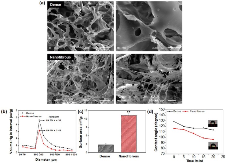
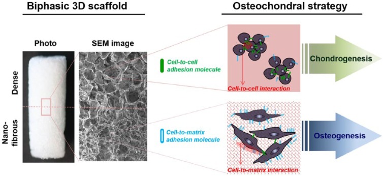
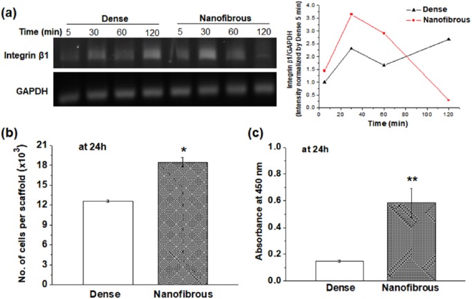
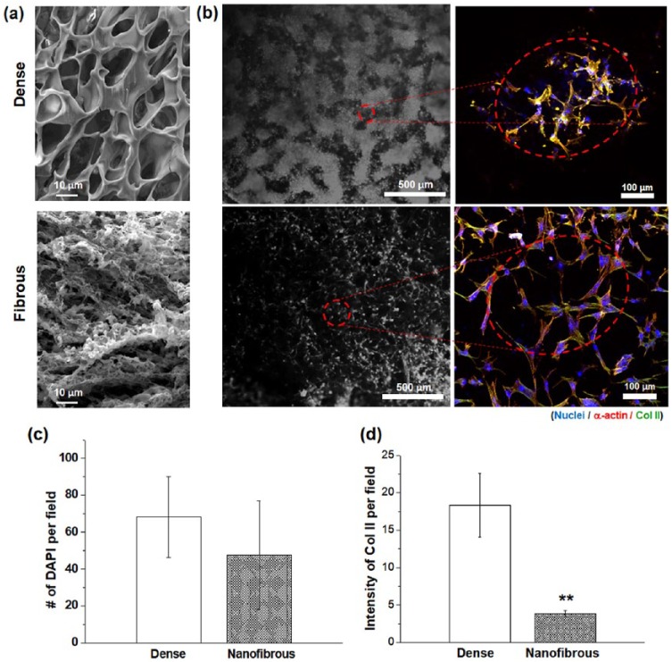
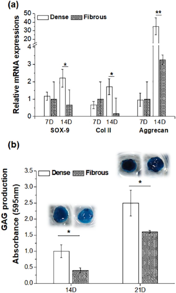
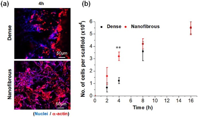
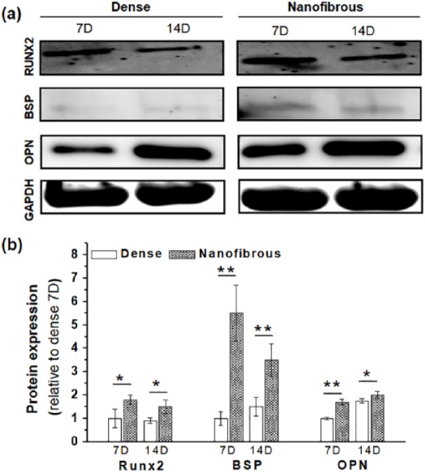
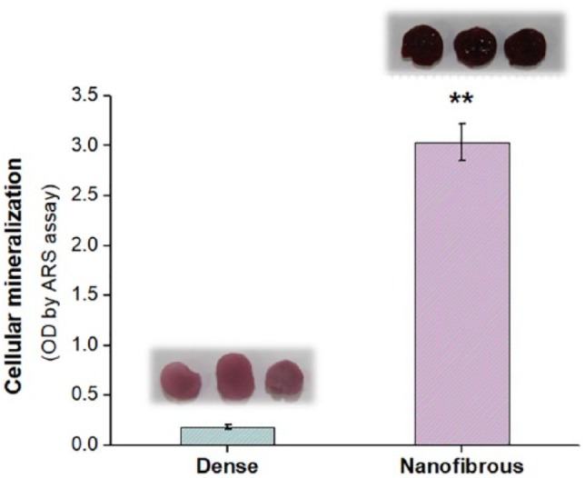
Bone/cartilage interfacial tissue engineering needs to satisfy the differential properties and architectures of the osteochondral region. Therefore, biphasic or multiphasic scaffolds that aim to mimic the gradient hierarchy are widely used. Here, we find that two differently structured (topographically) three-dimensional scaffolds, namely, "dense" and "nanofibrous" surfaces, show differential stimulation in osteo- and chondro-responses of cells. While the nanofibrous scaffolds accelerate the osteogenesis of mesenchymal stem cells, the dense scaffolds are better in preserving the phenotypes of chondrocytes. Two types of porous scaffolds, generated by a salt-leaching method combined with a phase-separation process using the poly(lactic acid) composition, had a similar level of porosity (90%) and pore size (150 μm). The major difference in the surface nanostructure led to substantial changes in the surface area and water hydrophilicity (nanofibrous ≫ dense); as a result, the nanofibrous scaffolds increased the cell-to-matrix adhesion of mesenchymal stem cells significantly while decreasing the cell-to-cell contracts. Importantly, the chondrocytes, when cultured on nanofibrous scaffolds, were prone to lose their phenotype, including reduced chondrogenic expressions (SOX-9, collagen type II, and Aggrecan) and glycosaminoglycan content, which was ascribed to the enhanced cell-matrix adhesion with reduced cell-cell contacts. On the contrary, the osteogenesis of mesenchymal stem cells was significantly accelerated by the improved cell-to-matrix adhesion, as evidenced in the enhanced osteogenic expressions (RUNX2, bone sialoprotein, and osteopontin) and cellular mineralization. Based on these findings, we consider that the dense scaffold is preferentially used for the chondral-part, whereas the nanofibrous structure is suitable for osteo-part, to provide an optimal biphasic matrix environment for osteochondral tissue engineering.
骨/软骨界面组织工程需要满足骨软骨区域不同的特性和结构。因此,旨在模拟梯度层次结构的双相或多相支架被广泛应用。在此,我们发现两种结构(拓扑结构)不同的三维支架,即“致密”和“纳米纤维”表面,对细胞的成骨和成软骨反应表现出不同的刺激作用。虽然纳米纤维支架能加速间充质干细胞的成骨作用,但致密支架在保留软骨细胞表型方面表现更佳。通过盐析法结合使用聚乳酸组合物的相分离过程制备的两种多孔支架,具有相似水平的孔隙率(约90%)和孔径(约150μm)。表面纳米结构的主要差异导致表面积和水亲水性发生显著变化(纳米纤维≫致密);结果,纳米纤维支架显著增加了间充质干细胞与基质的粘附力,同时减少了细胞间的收缩。重要的是,当软骨细胞在纳米纤维支架上培养时,容易失去其表型,包括软骨生成相关表达(SOX - 9、II型胶原蛋白和聚集蛋白聚糖)降低以及糖胺聚糖含量减少,这归因于细胞与基质粘附增强而细胞间接触减少。相反,间充质干细胞的成骨作用因细胞与基质粘附的改善而显著加速,这在增强的成骨相关表达(RUNX2、骨唾液蛋白和骨桥蛋白)以及细胞矿化中得到证实。基于这些发现,我们认为致密支架优先用于软骨部分,而纳米纤维结构适用于骨部分,以提供用于骨软骨组织工程的最佳双相基质环境。