von Bomhard Achim, Faust Joseph, Elsaesser Alexander F, Schwarz Silke, Pippich Katharina, Rotter Nicole
Department of Oral and Maxillofacial Surgery, The University Hospital Klinikum rechts der Isar, Munich, Germany.
Department of Oto-Rhino-Laryngology, Ulm University Medical Center, Ulm, Germany.
J Tissue Eng. 2017 Oct 6;8:2041731417732655. doi: 10.1177/2041731417732655. eCollection 2017 Jan-Dec.
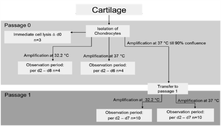
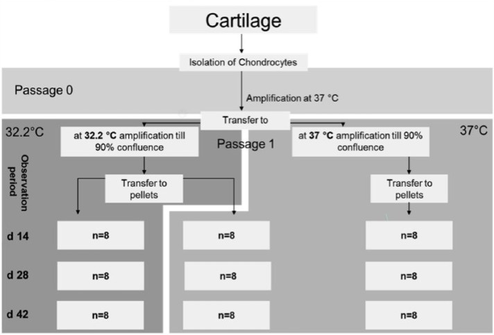
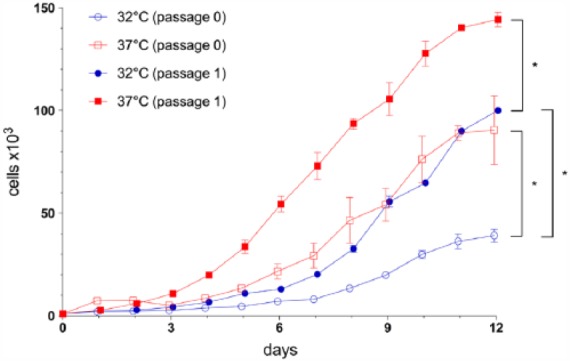
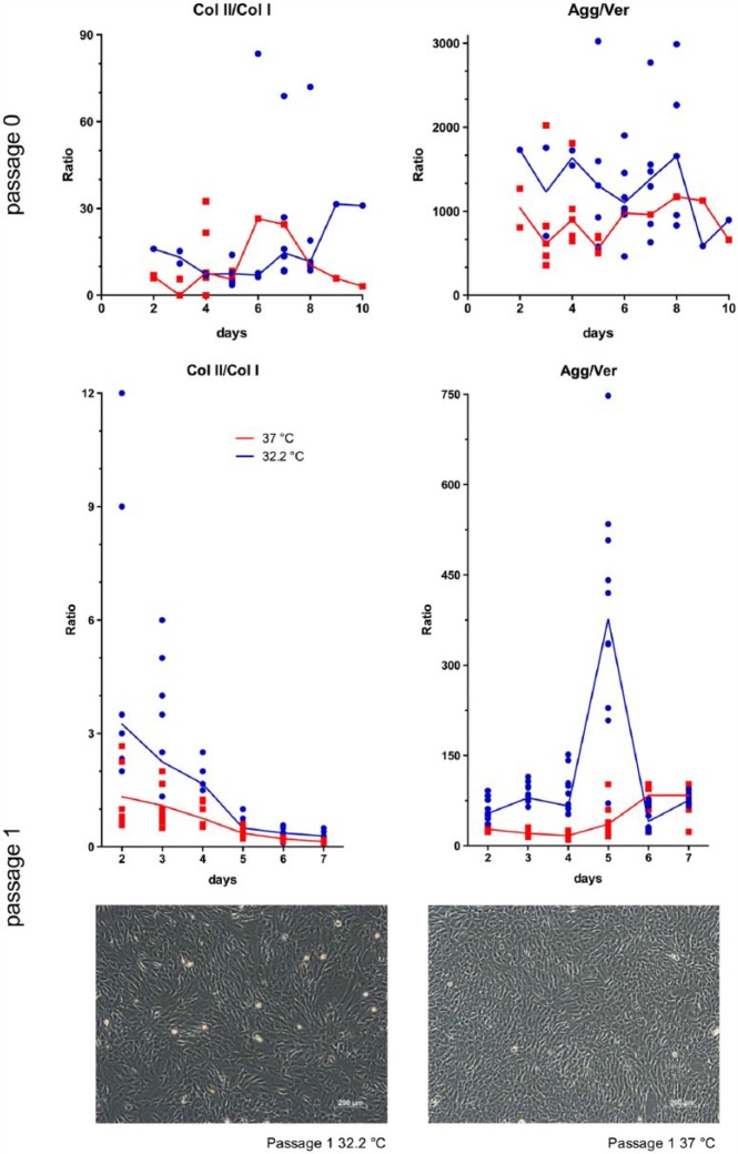
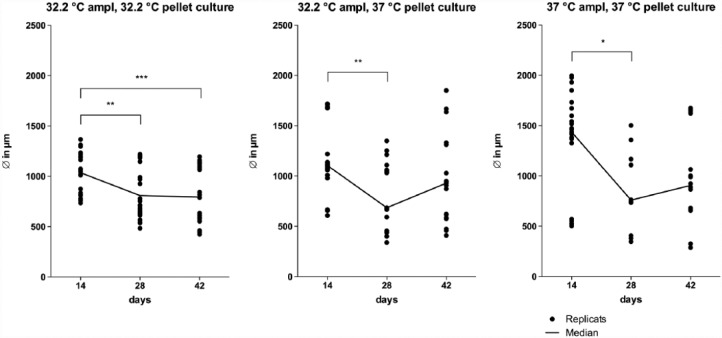
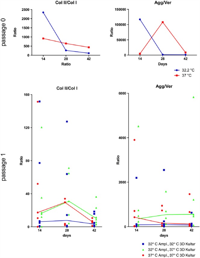
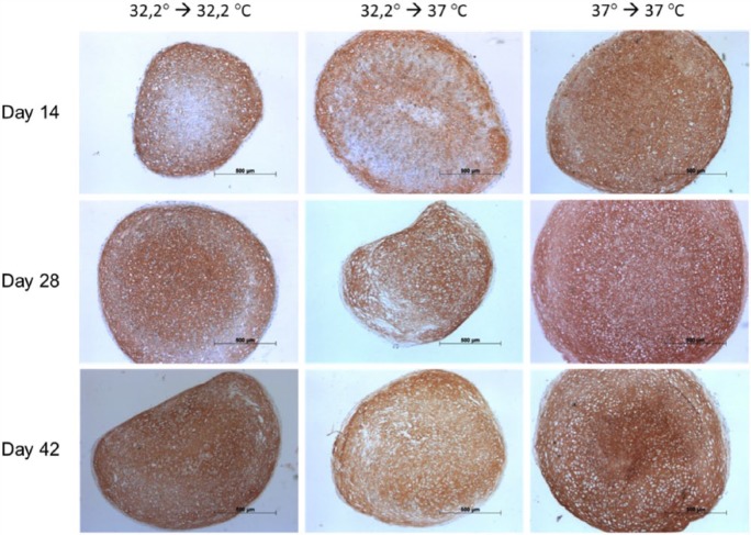
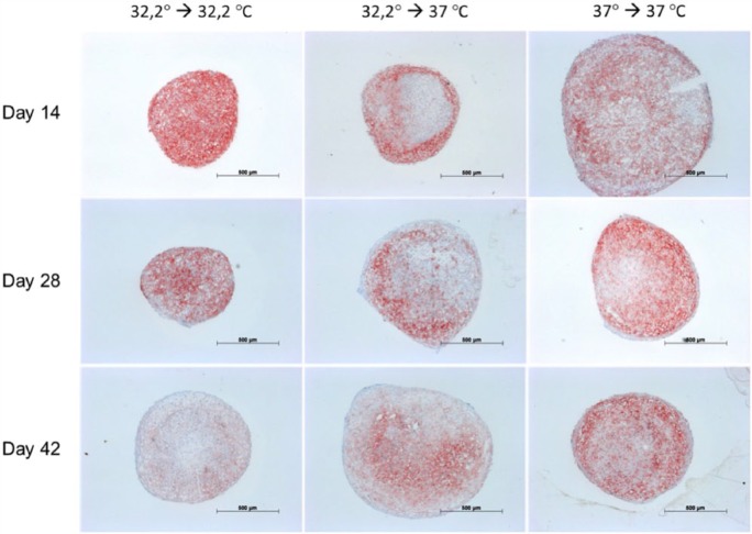
A critical limitation in the cultivation of cartilage for tissue engineering is the dedifferentiation in chondrocytes, mainly during in vitro amplification. Despite many previous studies investigating the influence of various conditions, no data exist concerning the effects of hypothermia. Our aim has been to influence chondrocyte dedifferentiation in vitro by hypothermic conditions. Chondrocytes were isolated from cartilage biopsies and seeded in monolayer and in three-dimensional pellet-cultures. Each cell culture was either performed at 32.2°C or 37°C during amplification. Additionally, the influence of the redifferentiation of chondrocytes in three-dimensional cell culture was examined at 32.2°C and 37°C after amplification at 32.2°C or 37°C. An 3-(4,5-dimethylthiazol-2-yl)-5-(3-carboxymethoxyphenyl)-2-(4-sulfophenyl)-2H-tetrazolium (MTS) assay was used to measure cell proliferation in monolayer, whereas the polymerase chain reaction and immunohistochemical and histological staining were used in three-dimensional pellet-cultures. Real-time polymerase chain reaction was employed to measure the relative expression of the target genes collagen II, collagen I, aggrecan and versican. Ratios were estimated between collagen II/collagen I and aggrecan/versican to evaluate differentiation. A higher value of these ratios indicated an advantageous status of differentiation. In monolayer, hypothermia at 32.2°C slowed down the proliferation rate of chondrocytes significantly, being up to two times lower at 32.2°C compared with culture at 37°C. Simultaneously, hypothermia in monolayer decelerated dedifferentiation. The ratio of aggrecan/versican was significantly higher at 32.2°C compared with that at 37°C. In three-dimensional pellet-culture, the chondrocytes redifferentiated at 32.2°C and at 37°C, and this process is more distinct at 37°C than at 32.2°C. Similar results were obtained for the ratios of collagen II/collagen I and aggrecan/versican and were supported by immunochemical and histological staining. Thus, hypothermic conditions for chondrocytes are mainly advantageous in monolayer culture. In three-dimensional pellet-culture, redifferentiation predominates at 37°C compared with at 32.2°C. In particular, the results from the monolayer cultures show potential in the avoidance of dedifferentiation.
组织工程中软骨培养的一个关键限制是软骨细胞的去分化,主要发生在体外扩增过程中。尽管此前有许多研究探讨了各种条件的影响,但关于低温影响的数据尚不存在。我们的目标是通过低温条件在体外影响软骨细胞的去分化。从软骨活检组织中分离软骨细胞,并接种于单层培养和三维微团培养中。在扩增过程中,每种细胞培养分别在32.2°C或37°C下进行。此外,在32.2°C或37°C扩增后,在32.2°C和37°C下检测三维细胞培养中软骨细胞再分化的影响。采用3-(4,5-二甲基噻唑-2)-5-(3-羧甲氧基苯基)-2-(4-磺基苯基)-2H-四唑(MTS)法检测单层培养中的细胞增殖,而在三维微团培养中采用聚合酶链反应、免疫组织化学和组织学染色。采用实时聚合酶链反应检测目标基因Ⅱ型胶原、Ⅰ型胶原、聚集蛋白聚糖和多功能蛋白聚糖的相对表达。计算Ⅱ型胶原/Ⅰ型胶原和聚集蛋白聚糖/多功能蛋白聚糖的比值以评估分化情况。这些比值越高表明分化状态越有利。在单层培养中,32.2°C的低温显著减慢了软骨细胞的增殖速率,与37°C培养相比,32.2°C时的增殖速率低至两倍。同时,单层培养中的低温减缓了去分化。32.2°C时聚集蛋白聚糖/多功能蛋白聚糖的比值显著高于37°C时。在三维微团培养中,软骨细胞在32.2°C和37°C下均发生再分化,且该过程在37°C时比在32.2°C时更明显。Ⅱ型胶原/Ⅰ型胶原和聚集蛋白聚糖/多功能蛋白聚糖的比值也得到了类似结果,并得到免疫化学和组织学染色的支持。因此,软骨细胞的低温条件主要有利于单层培养。在三维微团培养中,与32.2°C相比,37°C时再分化占主导。特别是,单层培养的结果显示出在避免去分化方面的潜力。