Neurovascular Research Group, Institute of Neuroscience, Newcastle University, Campus for Ageing and Vitality, Newcastle upon Tyne, NE4 5PL, UK.
Acta Neuropathol Commun. 2019 Feb 7;7(1):16. doi: 10.1186/s40478-019-0666-x.
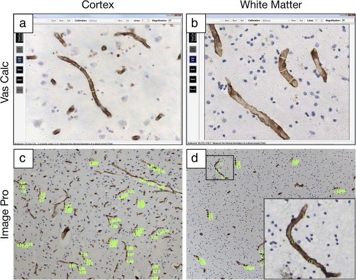
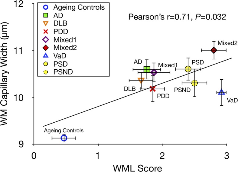
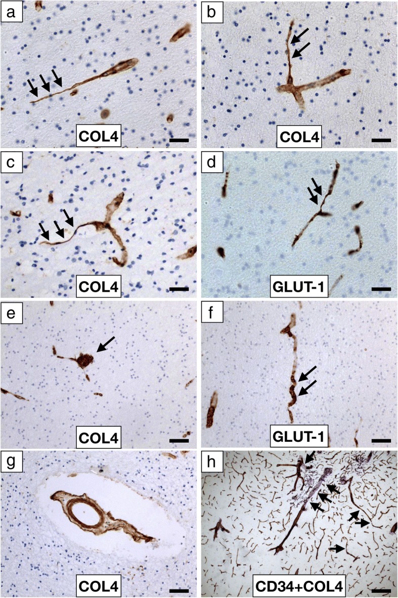
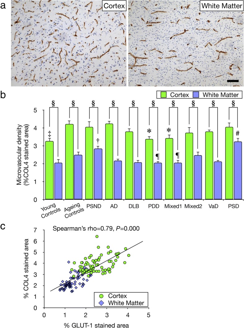
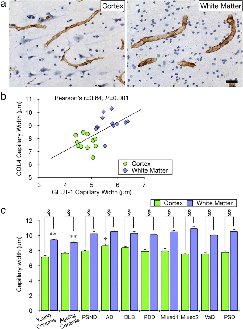
Previous studies suggest white matter (WM) integrity is vulnerable to chronic hypoperfusion during brain ageing. We assessed ~ 0.7 million capillary profiles in the frontal lobe WM across several dementias comprising Alzheimer's disease, dementia with Lewy bodies, Parkinson's disease with dementia, vascular dementia, mixed dementias, post-stroke dementia as well as post-stroke no dementia and similar age ageing and young controls without significant brain pathology. Standard histopathological methods were used to determine microvascular pathology and capillary width and densities in 153 subjects using markers of the basement membrane (collagen IV; COL4) and endothelium (glucose transporter-1; GLUT-1). Variable microvascular pathology including coiled, tortuous, collapsed and degenerated capillaries as well as occasional microaneurysms was present in all dementias. As expected, WM microvascular densities were 20-49% lower than in the overlying cortex. This differential in density between WM and cortex was clearly demonstrated by COL4, which was highly correlated with GLUT-1 densities (Spearman's rho = 0.79, P = 0.000). WM COL4 immunopositive microvascular densities were decreased by ~ 18% across the neurodegenerative dementias. However, we found WM COL4 densities were increased by ~ 57% in post-stroke dementia versus ageing and young controls and other dementias. Using three different methods to measure capillary diameters, we found WM capillaries to be significantly wider by 19-45% compared to those in overlying neocortex apparent with both COL4 and GLUT-1. Remarkably, WM capillary widths were increased by ~ 20% across all dementias compared to ageing and young controls (P < 0.01). We also noted mean WM pathology scores incorporating myelin loss, arteriolosclerosis and perivascular spacing were correlated with COL4 immunopositive capillary widths (Pearson's r = 0.71, P = 0.032). Our key finding indicates that WM capillaries are wider compared to those in the overlying neocortex in controls but they dilate further during dementia pathogenesis. We suggest capillaries undergo restructuring in the deep WM in different dementias. This reflects compensatory changes to retain WM perfusion and integrity during hypoperfusive states in ageing-related dementias.
先前的研究表明,在大脑老化过程中,慢性低灌注会使白质(WM)完整性受损。我们评估了包括阿尔茨海默病、路易体痴呆、帕金森病伴痴呆、血管性痴呆、混合性痴呆、中风后痴呆以及中风后无脑病和相似年龄的正常老化和年轻对照组在内的几种痴呆症中额叶 WM 中的约 70 万个毛细血管形态。使用基底膜标志物(IV 型胶原蛋白;COL4)和内皮标志物(葡萄糖转运蛋白-1;GLUT-1),对 153 名受试者的微血管病理和毛细血管宽度及密度进行了标准组织病理学方法的检测。在所有痴呆症中都存在可变的微血管病理,包括卷曲、扭曲、塌陷和退化的毛细血管以及偶尔的微动脉瘤。正如预期的那样,WM 微血管密度比皮层低 20-49%。COL4 清楚地证明了 WM 和皮层之间的这种密度差异,COL4 与 GLUT-1 密度高度相关(Spearman's rho=0.79,P=0.000)。在神经退行性痴呆症中,WM COL4 免疫阳性微血管密度降低了约 18%。然而,我们发现与正常老化和年轻对照组以及其他痴呆症相比,中风后痴呆症的 WM COL4 密度增加了约 57%。使用三种不同的方法测量毛细血管直径,我们发现 WM 毛细血管比皮层中的毛细血管宽 19-45%,这一点在 COL4 和 GLUT-1 中都很明显。值得注意的是,与正常老化和年轻对照组相比,所有痴呆症的 WM 毛细血管宽度增加了约 20%(P<0.01)。我们还注意到,纳入髓鞘丢失、小动脉硬化和血管周围间距的平均 WM 病理评分与 COL4 免疫阳性毛细血管宽度相关(Pearson's r=0.71,P=0.032)。我们的主要发现表明,与对照组皮层中的毛细血管相比,WM 毛细血管更宽,但在痴呆症发病机制中进一步扩张。我们认为在不同的痴呆症中,WM 毛细血管在深部 WM 中发生了重构。这反映了在与老化相关的痴呆症中的低灌注状态下,为保持 WM 灌注和完整性而发生的代偿性改变。