Neurovascular Research Group, Translational and Clinical Research Institute, Newcastle University, Campus for Ageing and Vitality, Newcastle Upon Tyne, NE4 5PL, UK.
Acta Neuropathol Commun. 2021 Aug 2;9(1):130. doi: 10.1186/s40478-021-01230-6.
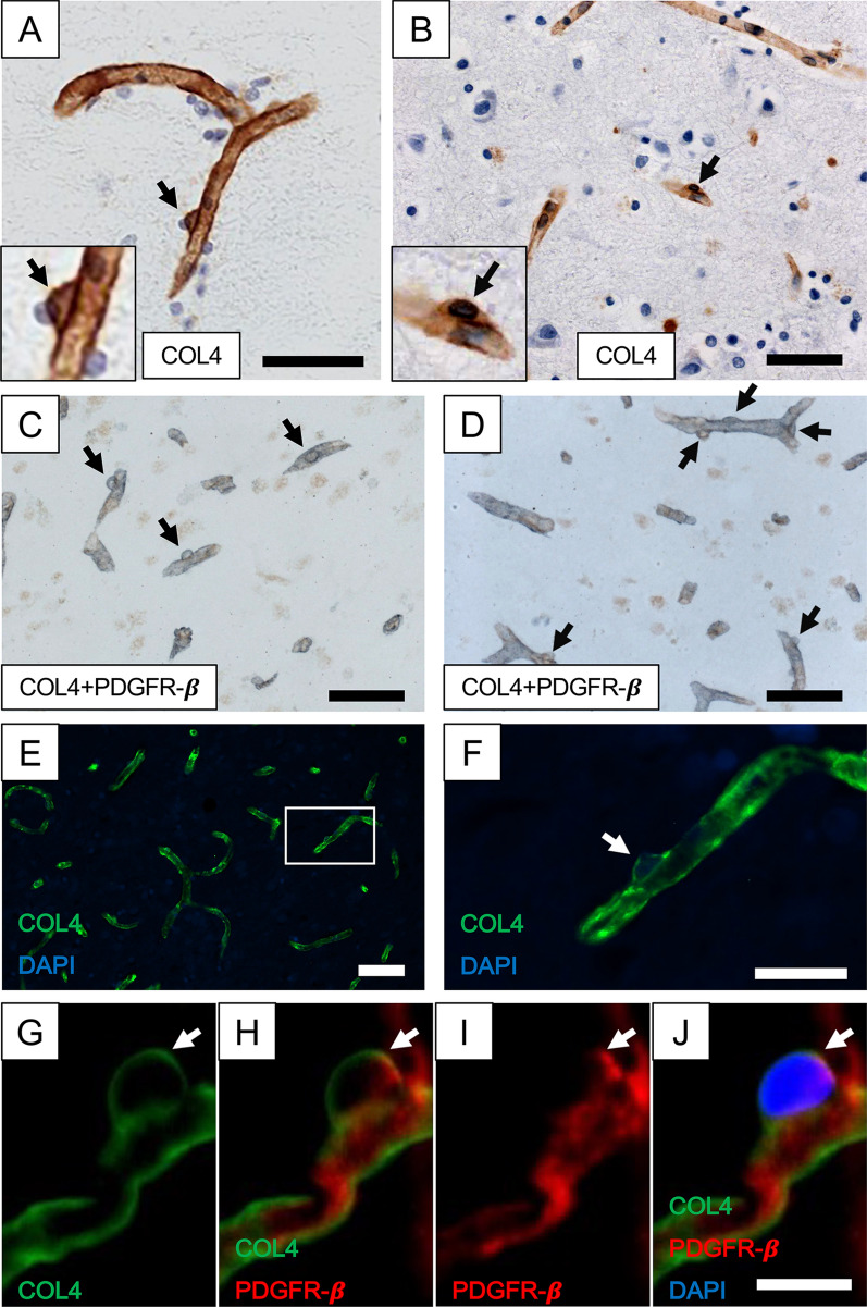
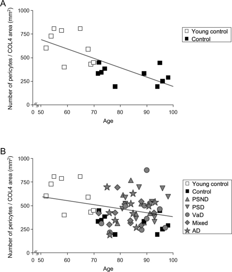
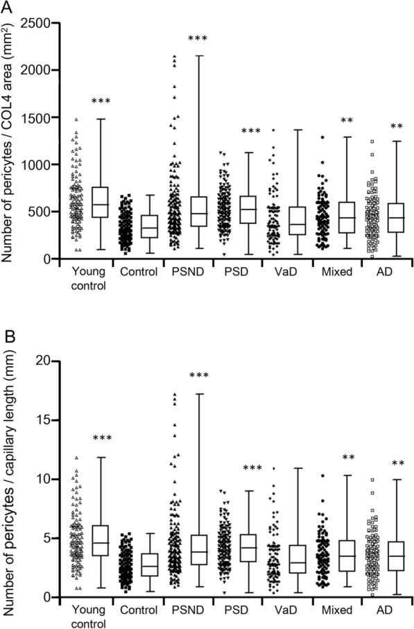
Cerebral pericytes are an integral component of the neurovascular unit, which governs the blood-brain barrier. There is paucity of knowledge on cortical pericytes across different dementias. We quantified cortical pericytes in capillaries in 124 post-mortem brains from subjects with post-stroke dementia (PSD), vascular dementia (VaD), Alzheimer's disease (AD) and AD-VaD (Mixed) and, post-stroke non-demented (PSND) stroke survivors as well as normal ageing controls. Collagen 4 (COL4)-positive nucleated pericyte soma were identified as protrusions on capillaries of the frontal cortex. The COL4-positive somata or nodule-like cell bodies were also verified by platelet derived growth factor receptor-β (PDGFR-β) immunohistochemistry. The mean (± SEM) pericyte somata in frontal cortical capillaries in normal young controls (46-65 years of age) was estimated as 5.2 ± 0.2 per mm capillary length. This number was reduced by 45% in older controls (> 78 years) to 2.9 ± 0.1 per mm capillary length (P < 0.001). We further found that the numbers of pericyte cell bodies per COL4 mm area or per mm capillary length were not decreased but rather preserved or increased in PSD, AD and Mixed dementia groups compared to similar age older controls (P < 0.01). Consistent with this, we noted that capillary length densities identified by the endothelial marker glucose transporter 1 or COL4 were not different across the dementias compared to older controls. There was a negative correlation with age (P < 0.001) suggesting fewer pericyte somata in older age, although the % COL4 immunoreactive capillary area was increased in older controls compared to young controls. Using a proven reliable method to quantify COL4-positive nucleated pericytes, our observations demonstrate ageing related loss but mostly preserved pericytes in the frontal cortex of vascular and AD dementias. We suggest there is differential regulation of capillary pericytes in the frontal lobe between the cortex and white matter in ageing-related dementias.
脑周细胞是神经血管单元的一个组成部分,它控制着血脑屏障。目前对不同痴呆症患者大脑皮层周细胞的了解甚少。我们对 124 例死后脑卒后痴呆(PSD)、血管性痴呆(VaD)、阿尔茨海默病(AD)和 AD-VaD(混合)以及卒中后非痴呆(PSND)幸存者以及正常老化对照者的大脑皮层毛细血管中的皮层周细胞进行了定量分析。用胶原蛋白 4(COL4)阳性有核周细胞体鉴定出在大脑皮层毛细血管上的突起。COL4 阳性的体或结节样细胞体也通过血小板衍生生长因子受体-β(PDGFR-β)免疫组织化学法得到了验证。正常年轻对照组(46-65 岁)大脑皮层毛细血管中周细胞体的平均(±SEM)估计为 5.2±0.2 个/mm 毛细血管长度。在年龄较大的对照组(>78 岁)中,这个数字减少了 45%,降至 2.9±0.1 个/mm 毛细血管长度(P<0.001)。我们还发现,与相似年龄的老年对照组相比,PSD、AD 和混合性痴呆组中 COL4 每毫米面积或每毫米毛细血管长度的周细胞体数量没有减少,而是保持或增加(P<0.01)。与此一致的是,我们注意到,与老年对照组相比,通过内皮标记葡萄糖转运蛋白 1 或 COL4 鉴定的毛细血管长度密度在不同痴呆症中没有差异。与年龄呈负相关(P<0.001),表明随着年龄的增长,周细胞体数量减少,尽管与年轻对照组相比,老年对照组的 COL4 免疫反应性毛细血管面积增加。使用一种经过验证的可靠方法来定量 COL4 阳性有核周细胞,我们的观察结果表明,在血管性和 AD 痴呆症的大脑皮层中,与年龄相关的周细胞损失,但大部分得到了保留。我们认为,在与年龄相关的痴呆症中,大脑皮层和白质之间的前脑叶毛细血管周细胞存在不同的调节。