Franchi Marco, Masola Valentina, Bellin Gloria, Onisto Maurizio, Karamanos Konstantinos-Athanasios, Piperigkou Zoi
Department for Life Quality Studies, University of Bologna, 47100 Rimini, Italy.
Renal Unit, Department of Medicine, University Hospital of Verona, 37100 Verona, Italy.
J Clin Med. 2019 Feb 7;8(2):213. doi: 10.3390/jcm8020213.
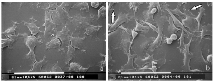
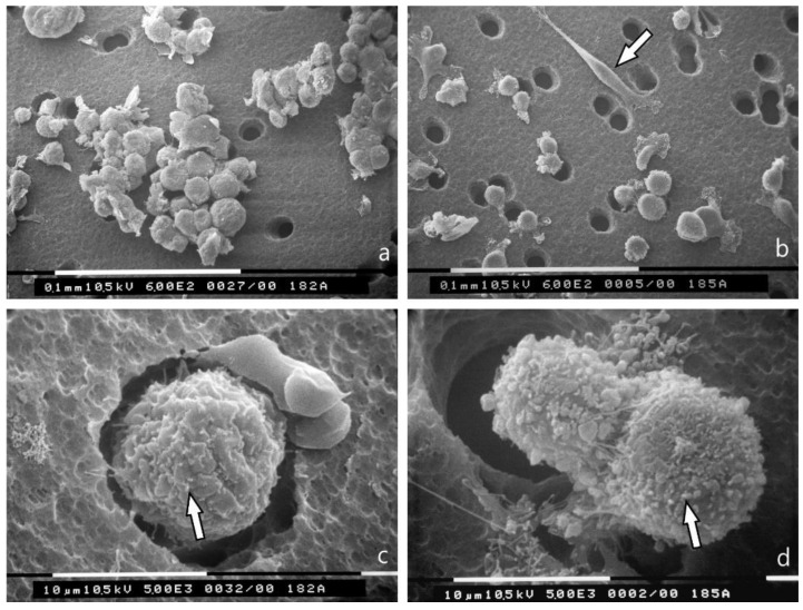
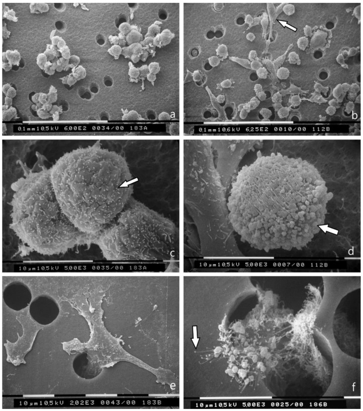
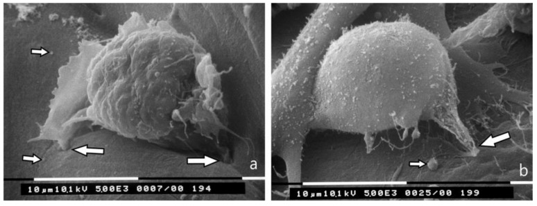
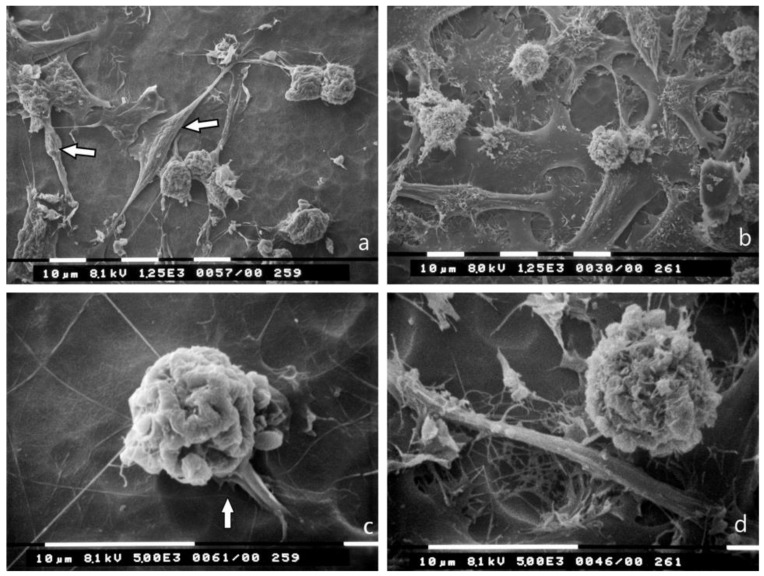
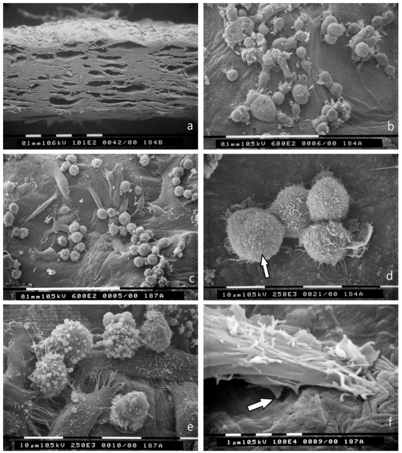
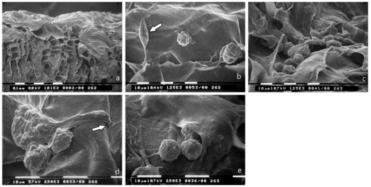
Interactions of cancer cells with matrix macromolecules of the surrounding tumor stroma are critical to mediate invasion and metastasis. In this study, we reproduced the collagen mechanical barriers in vitro (i.e., basement membrane, lamina propria under basement membrane, and deeper bundled collagen fibers with different array). These were used in 3D cell cultures to define their effects on morphology and behavior of breast cancer cells with different metastatic potential (MCF-7 and MDA-MB-231) using scanning electron microscope (SEM). We demonstrated that breast cancer cells cultured in 2D and 3D cultures on different collagen substrates show different morphologies: i) a globular/spherical shape, ii) a flattened polygonal shape, and iii) elongated/fusiform and spindle-like shapes. The distribution of different cell shapes changed with the distinct collagen fiber/fibril physical array and size. Dense collagen fibers, parallel to the culture plane, do not allow the invasion of MCF-7 and MDA-MB-231 cells, which, however, show increases of microvilli and microvesicles, respectively. These novel data highlight the regulatory role of different fibrillar collagen arrays in modifying breast cancer cell shape, inducing epithelial-to-mesenchymal transition, changing matrix composition and modulating the production of extracellular vesicles. Further investigation utilizing this in vitro model will help to demonstrate the biological roles of matrix macromolecules in cancer cell invasion in vivo.
癌细胞与周围肿瘤基质的大分子相互作用对于介导侵袭和转移至关重要。在本研究中,我们在体外重现了胶原机械屏障(即基底膜、基底膜下方的固有层以及具有不同排列的更深层束状胶原纤维)。这些被用于三维细胞培养,以使用扫描电子显微镜(SEM)确定它们对具有不同转移潜能的乳腺癌细胞(MCF-7和MDA-MB-231)的形态和行为的影响。我们证明,在不同胶原底物上进行二维和三维培养的乳腺癌细胞呈现出不同的形态:i)球形/球状形态,ii)扁平多边形形态,以及iii)细长/梭形和纺锤状形态。不同细胞形态的分布随胶原纤维/原纤维的不同物理排列和大小而变化。与培养平面平行的致密胶原纤维不允许MCF-7和MDA-MB-231细胞侵袭,然而,这两种细胞分别显示出微绒毛和微泡的增加。这些新数据突出了不同纤维状胶原排列在改变乳腺癌细胞形状、诱导上皮-间质转化、改变基质组成以及调节细胞外囊泡产生方面的调节作用。利用这种体外模型进行进一步研究将有助于证明基质大分子在体内癌细胞侵袭中的生物学作用。