Kim Seong Ho, Kim In Tae, Choi Chul Young
Department of Ophthalmology, Kangbuk Samsung Hospital, Sungkyunkwan University School of Medicine, Seoul, Korea.
Korean J Ophthalmol. 2019 Feb;33(1):8-15. doi: 10.3341/kjo.2018.0093.
To investigate the clinical manifestations and properties of remnant particles in the subconjunctival space after high-frequency radio-wave electrosurgery for conjunctivochalasis.
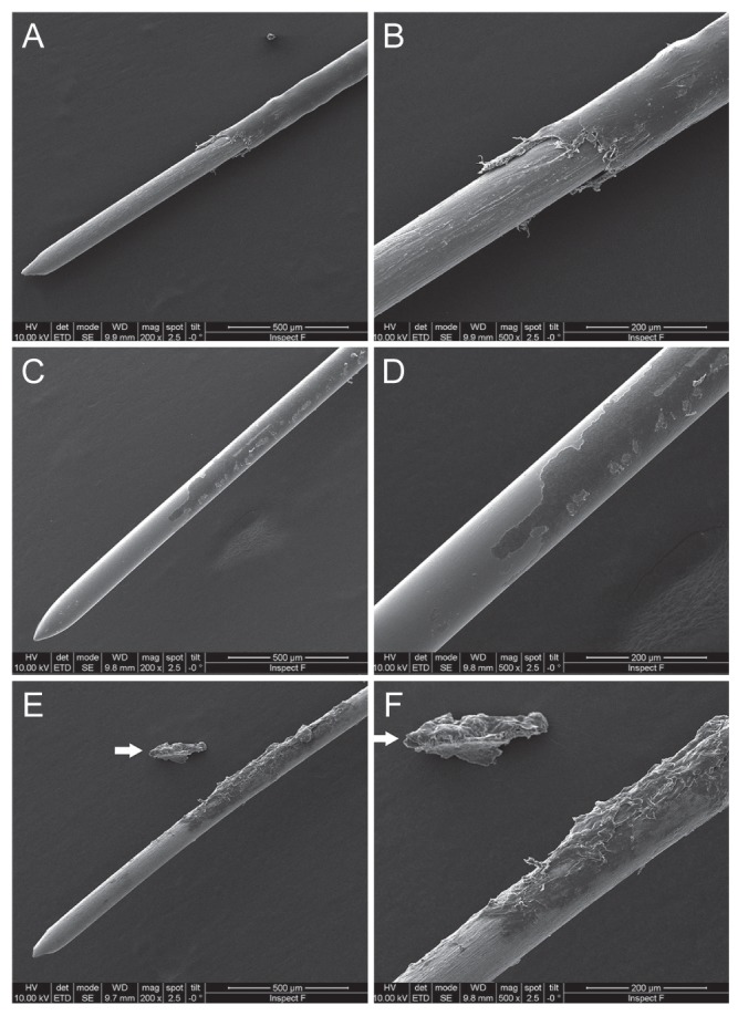
We performed a retrospective, observational case series with experimental imaging in nine eyes from eight patients who presented with small dark-gray lesions during follow-up after high-frequency radio-wave electrosurgery for conjunctivochalasis. General examination including slit-lamp examination and visual acuity testing was performed preoperatively and postoperatively. During follow-up, we evaluated remnant particles and any other complications including granuloma or conjunctival injection with slit-lamp photography and anterior optical coherence tomography. Coagulation tips were investigated with scanning electron microscope and energy dispersive X-ray spectroscopy to analyze the insulating electrode and assess changes to tips after repeated use.
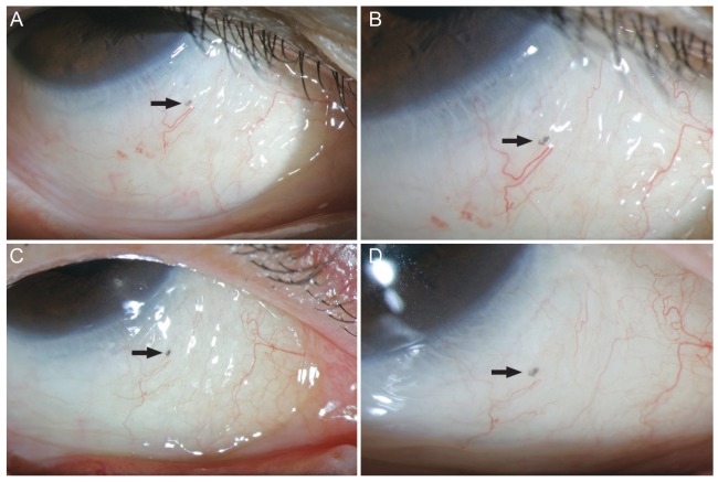
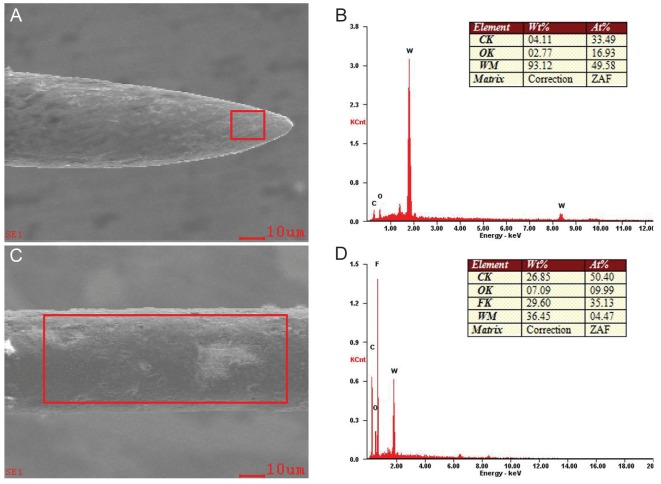
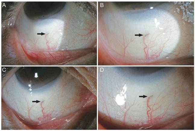
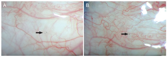
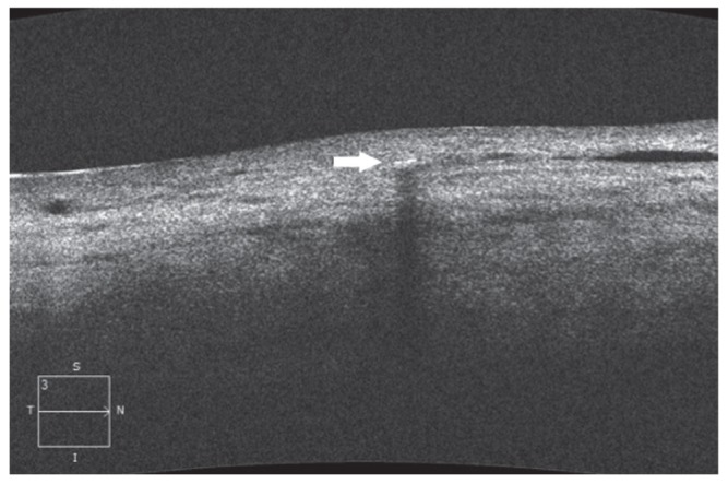
None of the patients included in this study experienced any change in visual acuity or major complications postoperatively. Small dark-gray lesions (0.3 to 0.5 mm in size) were observed in the inferior bulbar sub-conjunctival space in the location where high-frequency radio-wave electrosurgery had been performed. Cirrus high-definition optical coherence tomography images revealed focal hyper-reflection with a posterior shadow, suggesting foreign particles. Scanning electron microscopy and energy dispersive X-ray spectroscopy imaging analysis revealed peaks of carbon and fluorine complexes, consistent with the polytetrafluoroethylene coating on the electrode.
There were no instances of inflammatory reaction, particle migration, or major complications due to particles. Physicians should be aware of the possibility of remnant polytetrafluoroethylene particles in subconjunctival tissue when using insulated coagulation tips subjected to repeat sterilization.
探讨结膜松弛症高频电波电刀手术后结膜下间隙残留颗粒的临床表现及性质。
我们对8例患者的9只眼进行了一项回顾性观察病例系列研究,并进行了实验成像,这些患者在结膜松弛症高频电波电刀手术后随访期间出现小的深灰色病变。术前和术后进行了包括裂隙灯检查和视力测试在内的常规检查。在随访期间,我们通过裂隙灯摄影和眼前光学相干断层扫描评估残留颗粒及任何其他并发症,包括肉芽肿或结膜充血。用扫描电子显微镜和能量色散X射线光谱仪研究电凝头,以分析绝缘电极并评估重复使用后电凝头的变化。
本研究纳入的患者术后均未出现视力变化或重大并发症。在进行高频电波电刀手术的位置,在下睑结膜下间隙观察到小的深灰色病变(大小为0.3至0.5毫米)。Cirrus高清光学相干断层扫描图像显示局灶性高反射并伴有后部阴影,提示存在异物颗粒。扫描电子显微镜和能量色散X射线光谱成像分析显示碳和氟复合物的峰值,与电极上的聚四氟乙烯涂层一致。
未出现因颗粒引起的炎症反应、颗粒迁移或重大并发症的情况。使用经过反复灭菌的绝缘电凝头时,医生应意识到结膜下组织中可能残留聚四氟乙烯颗粒。