Al-Janabi Wissam, Krebs Renee, Arcila-Londono Ximena, Zaman Iram, Ahmad Bashiruddin K
Neurology Department, Henry Ford Health System, Detroit, MI, USA.
Eur J Case Rep Intern Med. 2018 Sep 27;5(9):000954. doi: 10.12890/2018_000954. eCollection 2018.
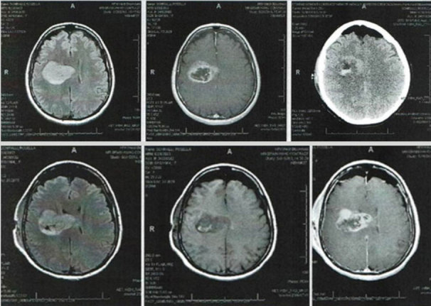
Glioblastoma multiforme (GBM) is a highly malignant glial tumour classified by the World Health Organization (WHO) as a stage IV astrocytoma. It varies in shape and size and can be cystic, vascular and necrotic. It often appears as a ring-enhancing lesion on magnetic resonance imaging (MRI). The most common symptoms of GBM, such as headache, vomiting and seizures, are due to increased intracranial pressure. The objective of this case report is to describe an atypical presentation of GBM.
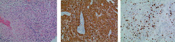
A 53-year-old woman of Italian origin presented with a 2-week history of lack of coordination in her hands and some difficulty in speech. Electromyography for assessment of her arms and cranial bulbar function was normal. However, 2 days later, the patient presented to the emergency department with progressive weakness in her left arm and leg as well as difficulty in speech. Mild left facial asymmetry was noted. A brain MRI revealed a right frontal mass. Stereotactic surgical resection was performed 2 days later, and biopsy confirmed the diagnosis of GBM. Although headache and other features of raised intracranial pressure are the most common initial symptoms of GBM, any atypical neurological or psychiatric presentation in an adult patient should raise suspicion for this tumour.
Careful analysis of an adult with atypical signs and symptoms along with thorough review of radiological tests will facilitate early diagnosis of dangerous tumours such as GBM.
An adult patient with symptoms that do not conform to a neurological condition should be investigated for a brain tumour.Careful history taking and examination are essential for reaching the correct diagnosis as soon as possible.Meticulous review of radiological images in order to detect subtle changes in brain anatomy is essential.
多形性胶质母细胞瘤(GBM)是一种高度恶性的胶质肿瘤,世界卫生组织(WHO)将其归类为IV级星形细胞瘤。其形状和大小各异,可呈囊性、血管性和坏死性。在磁共振成像(MRI)上,它常表现为环形强化病变。GBM最常见的症状,如头痛、呕吐和癫痫发作,是由颅内压升高引起的。本病例报告的目的是描述GBM的一种非典型表现。
一名53岁的意大利裔女性,有2周双手协调性差和言语略有困难的病史。评估其手臂和颅神经功能的肌电图检查正常。然而,2天后,患者因左臂和左腿进行性无力以及言语困难就诊于急诊科。发现有轻度左侧面部不对称。脑部MRI显示右额叶有肿块。2天后进行了立体定向手术切除,活检确诊为GBM。虽然头痛和其他颅内压升高的特征是GBM最常见的初始症状,但成年患者出现任何非典型的神经或精神表现都应怀疑患有这种肿瘤。
对有非典型体征和症状的成年人进行仔细分析,并对影像学检查进行全面复查,将有助于早期诊断GBM等危险肿瘤。
对于有不符合神经系统疾病症状的成年患者,应进行脑部肿瘤的检查。仔细询问病史和进行检查对于尽快做出正确诊断至关重要。细致复查影像学图像以检测脑部解剖结构的细微变化至关重要。