• 文献检索
  • 文档翻译
  • 深度研究
  • 学术资讯
  • Suppr Zotero 插件Zotero 插件
  • 邀请有礼
  • 套餐&价格
  • 历史记录
应用&插件
Suppr Zotero 插件Zotero 插件浏览器插件Mac 客户端Windows 客户端微信小程序
定价
高级版会员购买积分包购买API积分包
服务
文献检索文档翻译深度研究API 文档MCP 服务
关于我们
关于 Suppr公司介绍联系我们用户协议隐私条款
关注我们

Suppr 超能文献

核心技术专利:CN118964589B侵权必究
粤ICP备2023148730 号-1Suppr @ 2026

文献检索

告别复杂PubMed语法,用中文像聊天一样搜索,搜遍4000万医学文献。AI智能推荐,让科研检索更轻松。

立即免费搜索

文件翻译

保留排版,准确专业,支持PDF/Word/PPT等文件格式,支持 12+语言互译。

免费翻译文档

深度研究

AI帮你快速写综述,25分钟生成高质量综述,智能提取关键信息,辅助科研写作。

立即免费体验

脂质-蛋白质相互作用的新多样性。

Emerging Diversity in Lipid-Protein Interactions.

机构信息

Centre for Molecular Simulation and Department of Biological Sciences , University of Calgary , 2500 University Drive NW , Calgary , Alberta T2N 1N4 , Canada.

Groningen Biomolecular Sciences and Biotechnology Institute and Zernike Institute for Advanced Materials , University of Groningen , Nijenborgh 7 , 9747 AG Groningen , The Netherlands.

出版信息

Chem Rev. 2019 May 8;119(9):5775-5848. doi: 10.1021/acs.chemrev.8b00451. Epub 2019 Feb 13.

DOI:10.1021/acs.chemrev.8b00451
PMID:30758191
原文链接:https://pmc.ncbi.nlm.nih.gov/articles/PMC6509647/
Abstract

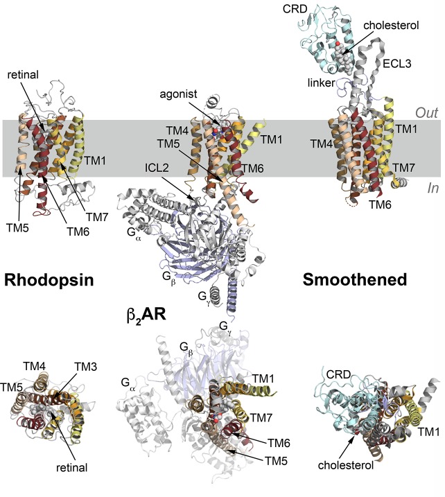
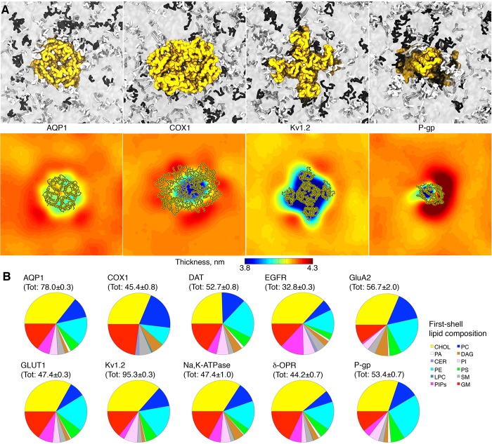
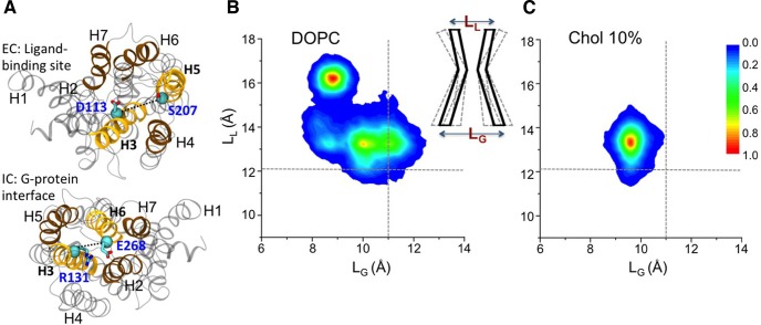
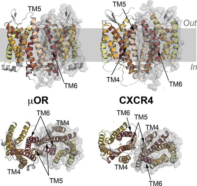
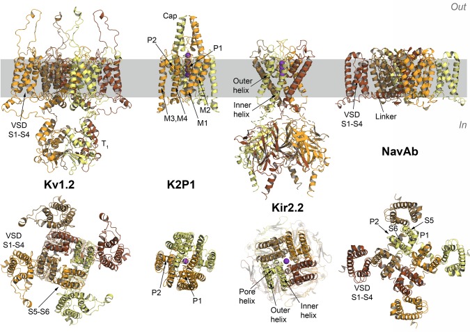
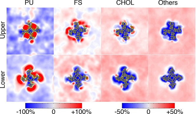
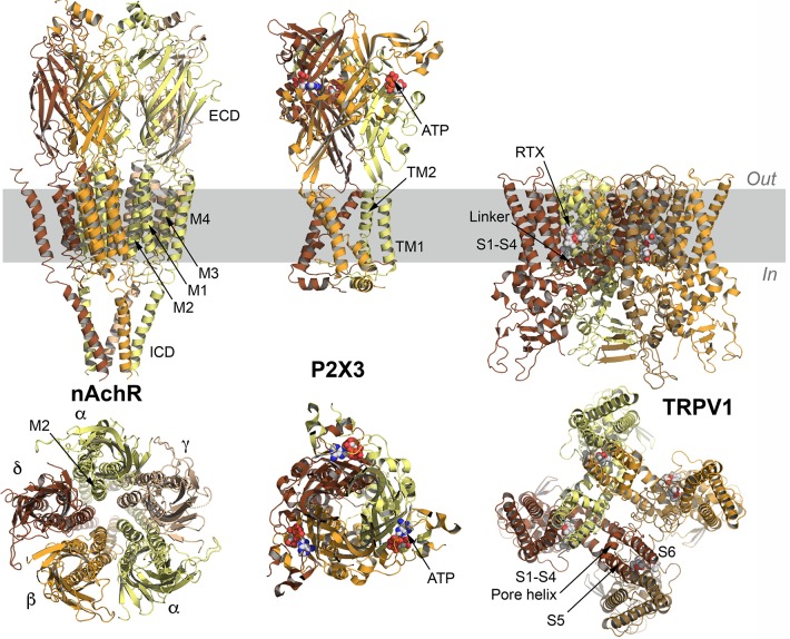
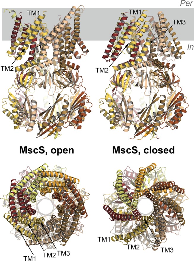
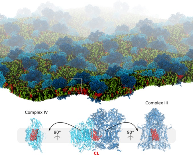
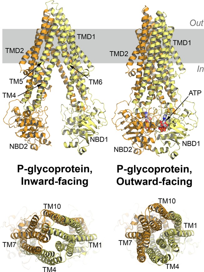
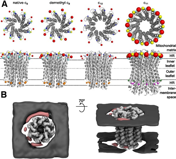
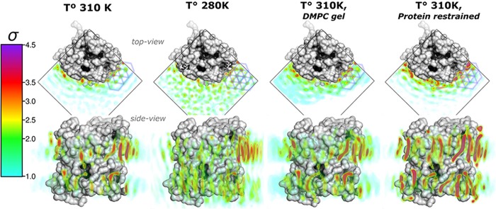
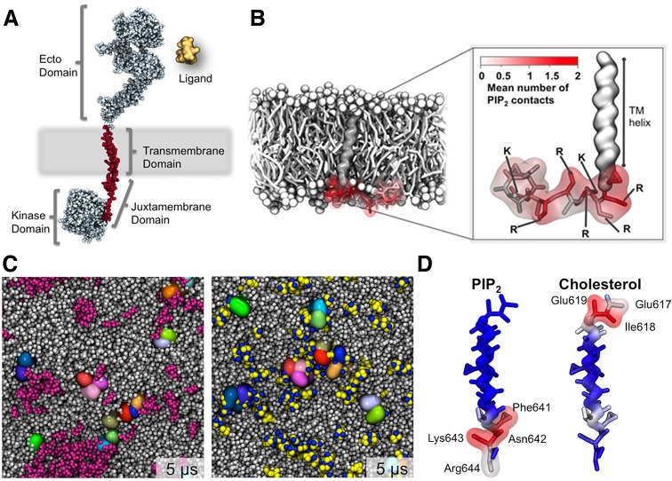
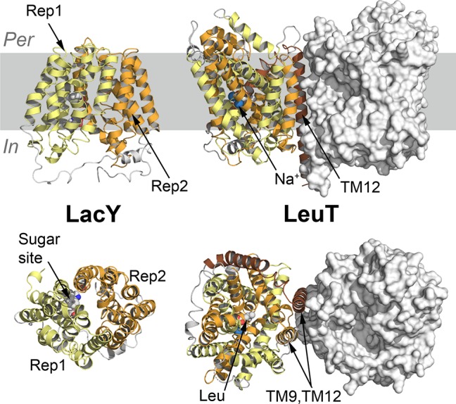
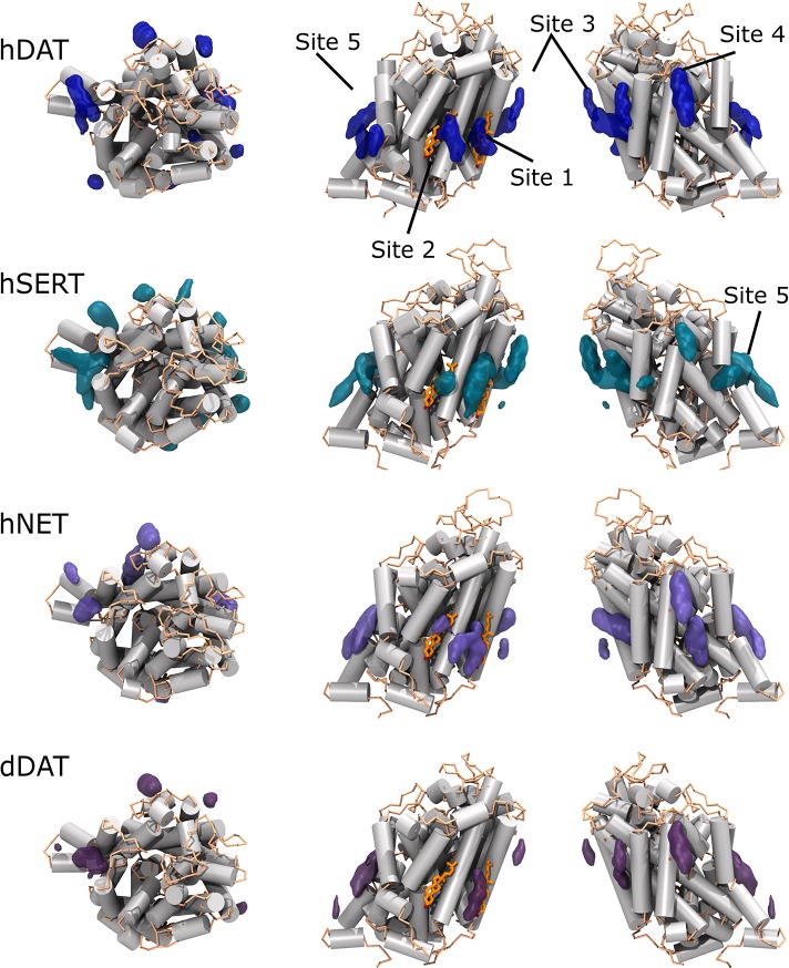
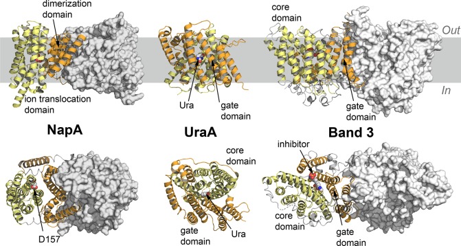
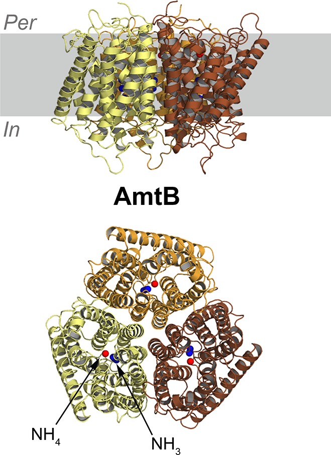
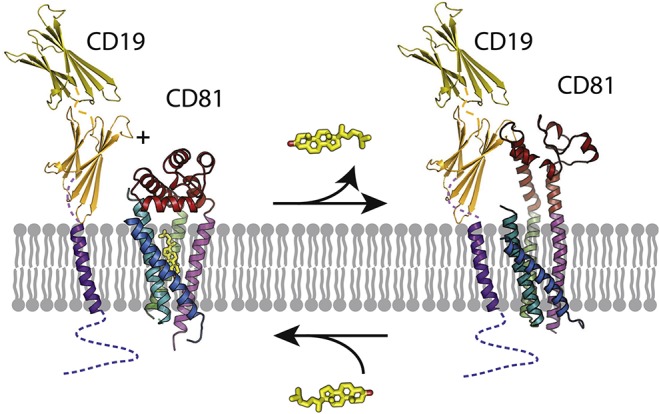
Membrane lipids interact with proteins in a variety of ways, ranging from providing a stable membrane environment for proteins to being embedded in to detailed roles in complicated and well-regulated protein functions. Experimental and computational advances are converging in a rapidly expanding research area of lipid-protein interactions. Experimentally, the database of high-resolution membrane protein structures is growing, as are capabilities to identify the complex lipid composition of different membranes, to probe the challenging time and length scales of lipid-protein interactions, and to link lipid-protein interactions to protein function in a variety of proteins. Computationally, more accurate membrane models and more powerful computers now enable a detailed look at lipid-protein interactions and increasing overlap with experimental observations for validation and joint interpretation of simulation and experiment. Here we review papers that use computational approaches to study detailed lipid-protein interactions, together with brief experimental and physiological contexts, aiming at comprehensive coverage of simulation papers in the last five years. Overall, a complex picture of lipid-protein interactions emerges, through a range of mechanisms including modulation of the physical properties of the lipid environment, detailed chemical interactions between lipids and proteins, and key functional roles of very specific lipids binding to well-defined binding sites on proteins. Computationally, despite important limitations, molecular dynamics simulations with current computer power and theoretical models are now in an excellent position to answer detailed questions about lipid-protein interactions.

摘要

膜脂以多种方式与蛋白质相互作用,从为蛋白质提供稳定的膜环境到在复杂且受到良好调节的蛋白质功能中发挥详细作用。实验和计算方面的进展正在脂质-蛋白质相互作用这一快速发展的研究领域中汇聚。在实验方面,高分辨率膜蛋白结构的数据库不断扩大,同时也具备了识别不同膜中复杂脂质组成、探测脂质-蛋白质相互作用具有挑战性的时间和长度尺度、以及将脂质-蛋白质相互作用与各种蛋白质的功能联系起来的能力。在计算方面,更精确的膜模型和更强大的计算机现在能够详细研究脂质-蛋白质相互作用,并与实验观察结果进行重叠,以验证和联合解释模拟和实验。在这里,我们综述了使用计算方法研究详细的脂质-蛋白质相互作用的论文,并简要介绍了实验和生理背景,旨在全面涵盖过去五年中模拟论文。总的来说,通过一系列机制,包括调节脂质环境的物理性质、脂质和蛋白质之间的详细化学相互作用以及非常特定的脂质与蛋白质上特定结合位点的关键功能作用,出现了一幅复杂的脂质-蛋白质相互作用图景。在计算方面,尽管存在重要限制,但目前的计算机能力和理论模型的分子动力学模拟现在处于回答有关脂质-蛋白质相互作用的详细问题的极佳位置。

https://cdn.ncbi.nlm.nih.gov/pmc/blobs/f080/6509647/e992cf19bc7d/cr-2018-00451c_0031.jpg
https://cdn.ncbi.nlm.nih.gov/pmc/blobs/f080/6509647/b1e99957e89e/cr-2018-00451c_0001.jpg
https://cdn.ncbi.nlm.nih.gov/pmc/blobs/f080/6509647/b8f4e1bac284/cr-2018-00451c_0002.jpg
https://cdn.ncbi.nlm.nih.gov/pmc/blobs/f080/6509647/ad890e02dae4/cr-2018-00451c_0003.jpg
https://cdn.ncbi.nlm.nih.gov/pmc/blobs/f080/6509647/ec5fb035c3ca/cr-2018-00451c_0004.jpg
https://cdn.ncbi.nlm.nih.gov/pmc/blobs/f080/6509647/953ed8d2f8cc/cr-2018-00451c_0005.jpg
https://cdn.ncbi.nlm.nih.gov/pmc/blobs/f080/6509647/e980b9034e53/cr-2018-00451c_0006.jpg
https://cdn.ncbi.nlm.nih.gov/pmc/blobs/f080/6509647/4b431ede8067/cr-2018-00451c_0007.jpg
https://cdn.ncbi.nlm.nih.gov/pmc/blobs/f080/6509647/1f05091c5185/cr-2018-00451c_0008.jpg
https://cdn.ncbi.nlm.nih.gov/pmc/blobs/f080/6509647/10f4ce2f3501/cr-2018-00451c_0009.jpg
https://cdn.ncbi.nlm.nih.gov/pmc/blobs/f080/6509647/6efc3e3604cd/cr-2018-00451c_0010.jpg
https://cdn.ncbi.nlm.nih.gov/pmc/blobs/f080/6509647/6232beb92282/cr-2018-00451c_0011.jpg
https://cdn.ncbi.nlm.nih.gov/pmc/blobs/f080/6509647/53a23c54eb60/cr-2018-00451c_0012.jpg
https://cdn.ncbi.nlm.nih.gov/pmc/blobs/f080/6509647/0e1b8de63e49/cr-2018-00451c_0013.jpg
https://cdn.ncbi.nlm.nih.gov/pmc/blobs/f080/6509647/17daa137deeb/cr-2018-00451c_0014.jpg
https://cdn.ncbi.nlm.nih.gov/pmc/blobs/f080/6509647/918d3cd1a027/cr-2018-00451c_0015.jpg
https://cdn.ncbi.nlm.nih.gov/pmc/blobs/f080/6509647/f4bbfe5efb40/cr-2018-00451c_0016.jpg
https://cdn.ncbi.nlm.nih.gov/pmc/blobs/f080/6509647/c0c17626c375/cr-2018-00451c_0017.jpg
https://cdn.ncbi.nlm.nih.gov/pmc/blobs/f080/6509647/a0905a82723e/cr-2018-00451c_0018.jpg
https://cdn.ncbi.nlm.nih.gov/pmc/blobs/f080/6509647/2a6b58d474ff/cr-2018-00451c_0019.jpg
https://cdn.ncbi.nlm.nih.gov/pmc/blobs/f080/6509647/4fc3cc5f0ef9/cr-2018-00451c_0020.jpg
https://cdn.ncbi.nlm.nih.gov/pmc/blobs/f080/6509647/c8b751c8cdd6/cr-2018-00451c_0021.jpg
https://cdn.ncbi.nlm.nih.gov/pmc/blobs/f080/6509647/6cf0be74ccfc/cr-2018-00451c_0022.jpg
https://cdn.ncbi.nlm.nih.gov/pmc/blobs/f080/6509647/03c23b3b4b30/cr-2018-00451c_0023.jpg
https://cdn.ncbi.nlm.nih.gov/pmc/blobs/f080/6509647/c574dcda9df2/cr-2018-00451c_0024.jpg
https://cdn.ncbi.nlm.nih.gov/pmc/blobs/f080/6509647/6c3ec5769e9c/cr-2018-00451c_0025.jpg
https://cdn.ncbi.nlm.nih.gov/pmc/blobs/f080/6509647/30000f019f6c/cr-2018-00451c_0026.jpg
https://cdn.ncbi.nlm.nih.gov/pmc/blobs/f080/6509647/7ce5883b614f/cr-2018-00451c_0027.jpg
https://cdn.ncbi.nlm.nih.gov/pmc/blobs/f080/6509647/37f27a66e73b/cr-2018-00451c_0028.jpg
https://cdn.ncbi.nlm.nih.gov/pmc/blobs/f080/6509647/1cc25e8e01e9/cr-2018-00451c_0029.jpg
https://cdn.ncbi.nlm.nih.gov/pmc/blobs/f080/6509647/6050fcfde94c/cr-2018-00451c_0030.jpg
https://cdn.ncbi.nlm.nih.gov/pmc/blobs/f080/6509647/e992cf19bc7d/cr-2018-00451c_0031.jpg

相似文献

1
Emerging Diversity in Lipid-Protein Interactions.脂质-蛋白质相互作用的新多样性。
Chem Rev. 2019 May 8;119(9):5775-5848. doi: 10.1021/acs.chemrev.8b00451. Epub 2019 Feb 13.
2
Characterization of Lipid-Protein Interactions and Lipid-Mediated Modulation of Membrane Protein Function through Molecular Simulation.通过分子模拟研究脂质-蛋白相互作用及脂质对膜蛋白功能的调控。
Chem Rev. 2019 May 8;119(9):6086-6161. doi: 10.1021/acs.chemrev.8b00608. Epub 2019 Apr 12.
3
Microscopic view of lipids and their diverse biological functions.脂质的微观结构及其多样化的生物学功能。
Curr Opin Struct Biol. 2018 Aug;51:177-186. doi: 10.1016/j.sbi.2018.07.003. Epub 2018 Jul 23.
4
Protein crowding and lipid complexity influence the nanoscale dynamic organization of ion channels in cell membranes.蛋白质拥挤和脂质复杂性影响细胞膜中离子通道的纳米级动态组织。
Sci Rep. 2017 Nov 30;7(1):16647. doi: 10.1038/s41598-017-16865-6.
5
Membrane transporter dimerization driven by differential lipid solvation energetics of dissociated and associated states.膜转运蛋白二聚化由解离态和结合态的差异脂溶剂化能驱动。
Elife. 2021 Apr 7;10:e63288. doi: 10.7554/eLife.63288.
6
The importance of membrane defects-lessons from simulations.膜缺陷的重要性:模拟研究的启示。
Acc Chem Res. 2014 Aug 19;47(8):2244-51. doi: 10.1021/ar4002729. Epub 2014 Jun 3.
7
Computational Modeling of Realistic Cell Membranes.真实细胞膜的计算建模。
Chem Rev. 2019 May 8;119(9):6184-6226. doi: 10.1021/acs.chemrev.8b00460. Epub 2019 Jan 9.
8
Emerging roles for lipids in shaping membrane-protein function.脂质在塑造膜蛋白功能方面的新作用。
Nature. 2009 May 21;459(7245):379-85. doi: 10.1038/nature08147.
9
Organizing membrane-curving proteins: the emerging dynamical picture.组织膜弯曲蛋白:新兴的动力学图景。
Curr Opin Struct Biol. 2018 Aug;51:99-105. doi: 10.1016/j.sbi.2018.03.018. Epub 2018 Mar 30.
10
The electrical interplay between proteins and lipids in membranes.膜中蛋白质与脂质之间的电相互作用。
Biochim Biophys Acta. 2015 Sep;1848(9):1828-36. doi: 10.1016/j.bbamem.2015.03.017. Epub 2015 Mar 24.

引用本文的文献

1
Bilayer-Dependent Recognition of Docosahexaenoic Acid by the Transmembrane Domain of FATP3.FATP3跨膜结构域对二十二碳六烯酸的双层依赖性识别
J Membr Biol. 2025 Sep 15. doi: 10.1007/s00232-025-00361-4.
2
PIP2 regulation of TRPV4 channels: Binding sites and dynamic coupling.磷脂酰肌醇-4,5-二磷酸(PIP2)对瞬时受体电位香草酸亚型4(TRPV4)通道的调节:结合位点与动态偶联
Biophys J. 2025 Aug 6. doi: 10.1016/j.bpj.2025.08.006.
3
Advances in native cell membrane nanoparticles system.天然细胞膜纳米颗粒系统的研究进展

本文引用的文献

1
Computational Modeling of Realistic Cell Membranes.真实细胞膜的计算建模。
Chem Rev. 2019 May 8;119(9):6184-6226. doi: 10.1021/acs.chemrev.8b00460. Epub 2019 Jan 9.
2
Cholesterol Interaction Sites on the Transmembrane Domain of the Hedgehog Signal Transducer and Class F G Protein-Coupled Receptor Smoothened.胆固醇在 Hedgehog 信号转导跨膜域和 Class F G 蛋白偶联受体 Smoothened 上的相互作用位点。
Structure. 2019 Mar 5;27(3):549-559.e2. doi: 10.1016/j.str.2018.11.003. Epub 2018 Dec 27.
3
Atomistic and Coarse Grain Simulations of the Cell Envelope of Gram-Negative Bacteria: What Have We Learned?
Curr Opin Struct Biol. 2025 Aug 6;94:103130. doi: 10.1016/j.sbi.2025.103130.
4
Biophysical Consequences for Exposure of Model Cell Membranes to Perfluoroalkyl Substances.模型细胞膜暴露于全氟烷基物质的生物物理后果。
J Phys Chem B. 2025 Aug 7;129(31):7951-7963. doi: 10.1021/acs.jpcb.5c02472. Epub 2025 Jul 28.
5
Temperature-dependence of membrane protein-lipid interactions in membranes.膜中膜蛋白 - 脂质相互作用的温度依赖性。
Chem Commun (Camb). 2025 Jun 27. doi: 10.1039/d5cc01576f.
6
Inside the membrane: a closer look using elastic scattering techniques and friends.膜内部:运用弹性散射技术及相关方法的深入观察
Eur Biophys J. 2025 Jun 26. doi: 10.1007/s00249-025-01761-z.
7
Unique Structural Features Relate to Evolutionary Adaptation of Cytochrome P450 in the Abyssal Zone.独特的结构特征与深海区域细胞色素P450的进化适应性相关。
Int J Mol Sci. 2025 Jun 13;26(12):5689. doi: 10.3390/ijms26125689.
8
Nanodisc single-molecule pulldown to study lipid-protein interactions.利用纳米圆盘单分子下拉技术研究脂类-蛋白质相互作用。
J Lipid Res. 2025 Jul;66(7):100846. doi: 10.1016/j.jlr.2025.100846. Epub 2025 Jun 20.
9
Global and local effects in lipid-mediated interactions between peripheral and integral membrane proteins.外周膜蛋白与整合膜蛋白之间脂质介导相互作用中的全局和局部效应。
Front Mol Biosci. 2025 May 30;12:1605772. doi: 10.3389/fmolb.2025.1605772. eCollection 2025.
10
Biosynthesis of lanosterol in .羊毛甾醇在……中的生物合成
Synth Syst Biotechnol. 2025 May 13;10(3):993-1001. doi: 10.1016/j.synbio.2025.05.006. eCollection 2025 Sep.
革兰氏阴性菌细胞包膜的原子级和粗粒级模拟:我们学到了什么?
Acc Chem Res. 2019 Jan 15;52(1):180-188. doi: 10.1021/acs.accounts.8b00377. Epub 2018 Dec 18.
4
Determining the target of membrane sterols on voltage-gated potassium channels.确定电压门控钾通道上膜甾醇的靶标。
Biochim Biophys Acta Mol Cell Biol Lipids. 2019 Mar;1864(3):312-325. doi: 10.1016/j.bbalip.2018.12.006. Epub 2018 Dec 13.
5
Coarse-grained molecular dynamics of membrane semitoroidal pore formation in model lipid-peptide systems.模型脂质-肽系统中膜半管状孔形成的粗粒度分子动力学。
J Mol Graph Model. 2019 Mar;87:1-10. doi: 10.1016/j.jmgm.2018.11.003. Epub 2018 Nov 13.
6
Effect of late endosomal DOBMP lipid and traditional model lipids of electrophysiology on the anthrax toxin channel activity.晚期内体 DOBMP 脂质和电生理学传统模型脂质对炭疽毒素通道活性的影响。
Biochim Biophys Acta Biomembr. 2018 Nov;1860(11):2192-2203. doi: 10.1016/j.bbamem.2018.08.011. Epub 2018 Aug 23.
7
Direct protein-lipid interactions shape the conformational landscape of secondary transporters.直接的蛋白质-脂质相互作用塑造了次级转运蛋白的构象景观。
Nat Commun. 2018 Oct 8;9(1):4151. doi: 10.1038/s41467-018-06704-1.
8
The Role of Lipid Interactions in Simulations of the α-Hemolysin Ion-Channel-Forming Toxin.脂质相互作用在α-溶血素离子通道形成毒素模拟中的作用。
Biophys J. 2018 Nov 6;115(9):1720-1730. doi: 10.1016/j.bpj.2018.09.009. Epub 2018 Sep 18.
9
Allosteric Role of Substrate Occupancy Toward the Alignment of P-glycoprotein Nucleotide Binding Domains.变构底物占据对 P-糖蛋白核苷酸结合域排列的作用。
Sci Rep. 2018 Oct 2;8(1):14643. doi: 10.1038/s41598-018-32815-2.
10
Distribution of mechanical stress in the Escherichia coli cell envelope.大肠杆菌细胞包膜中机械应力的分布。
Biochim Biophys Acta Biomembr. 2018 Dec;1860(12):2566-2575. doi: 10.1016/j.bbamem.2018.09.020. Epub 2018 Sep 29.