Wang Yang, Bao De-Jun, Xu Bin, Cheng Chuan-Dong, Dong Yong-Fei, Wei Xiang-Pin, Niu Chao-Shi
Department of Neurosurgery, First Affiliated Hospital of USTC, Division of Life Sciences and Medicine, University of Science and Technology of China, Hefei, Anhui Province, China.
Anhui Medical University Auhui Province Medical Genetic Center, Hefei, Anhui Province, China.
Neural Regen Res. 2019 Jun;14(6):1013-1024. doi: 10.4103/1673-5374.250620.
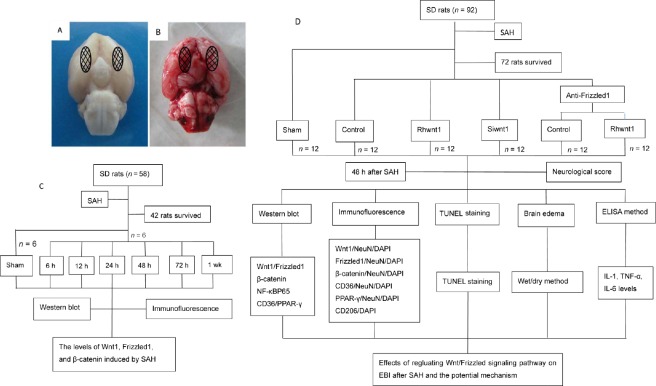
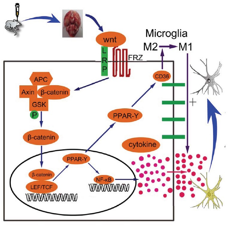
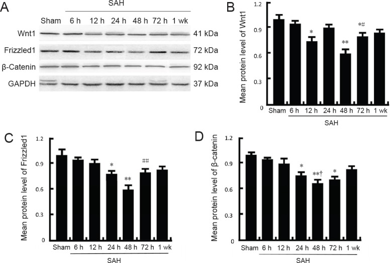
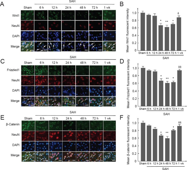
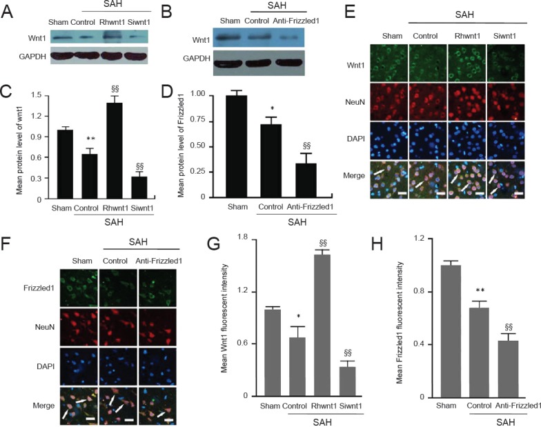
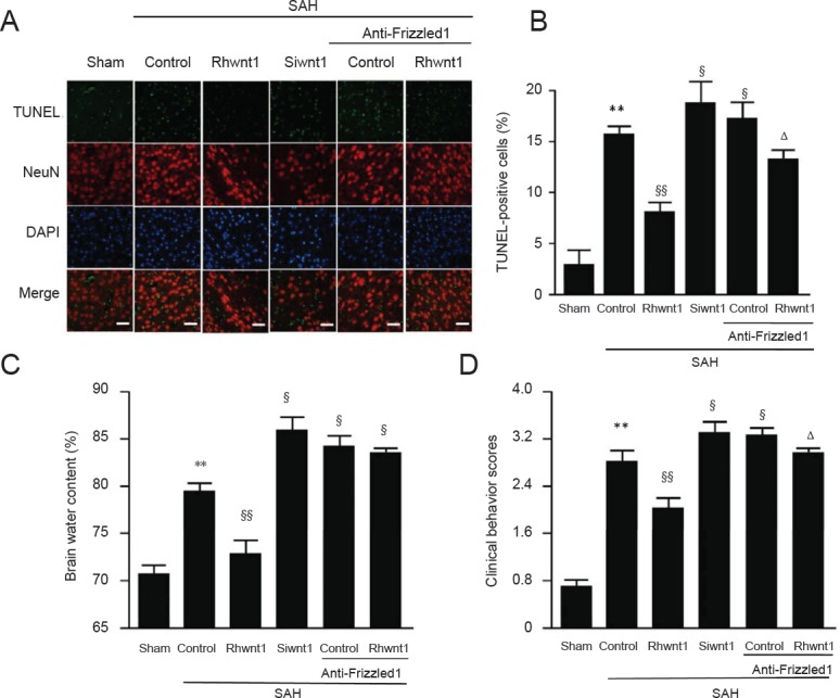
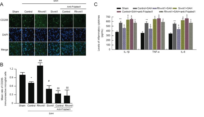
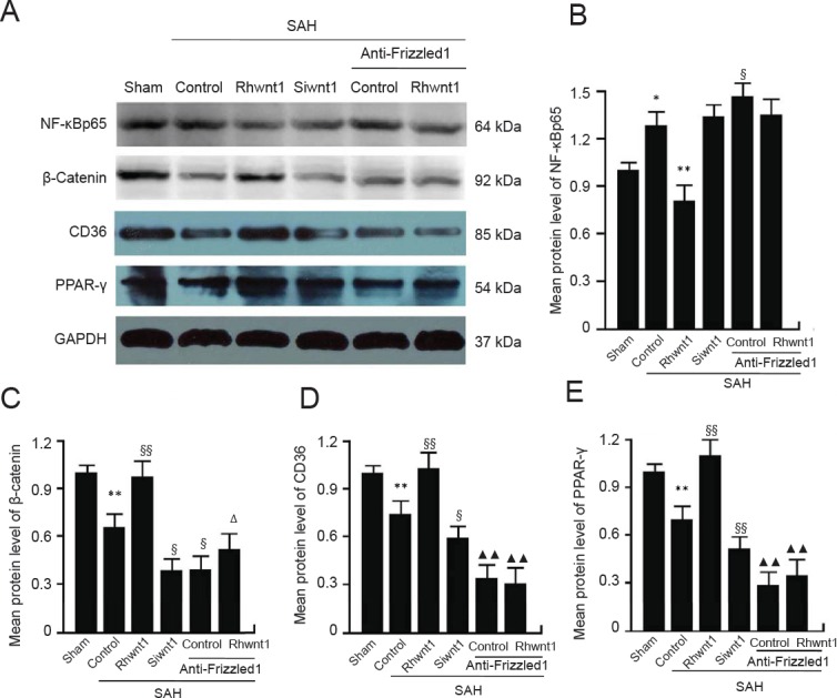
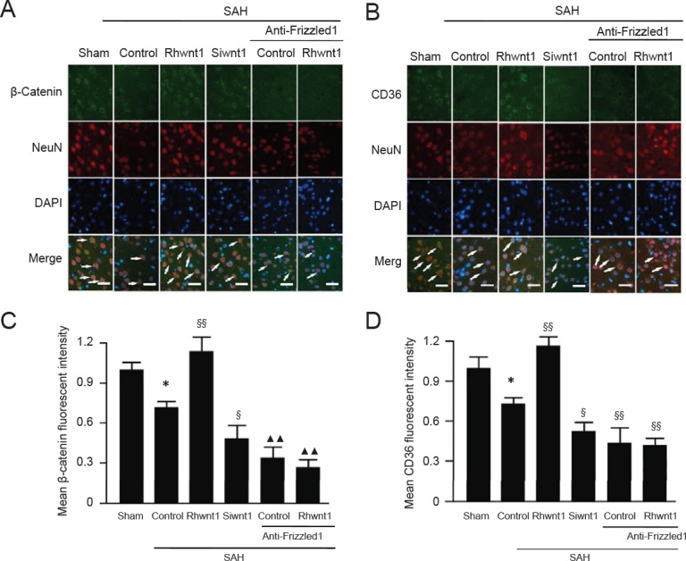
The Wnt/Frizzled signaling pathway participates in many inflammation-linked diseases. However, the inflammatory response mediated by the Wnt/Frizzled signaling pathway in experimental subarachnoid hemorrhage has not been thoroughly investigated. Consequently, in this study, we examined the potential role of the Wnt/Frizzled signaling pathway in early brain injury in rat models of subarachnoid hemorrhage. Simultaneously, possible neuroprotective mechanisms were also investigated. Experimental subarachnoid hemorrhage rat models were induced by injecting autologous blood into the prechiasmatic cistern. Experiment 1 was designed to examine expression of the Wnt/Frizzled signaling pathway in early brain injury induced by subarachnoid hemorrhage. In total, 42 adult rats were divided into sham (injection of equivalent volume of saline), 6-, 12-, 24-, 48-, 72-hour, and 1-week subarachnoid hemorrhage groups. Experiment 2 was designed to examine neuroprotective mechanisms of the Wnt/Frizzled signaling pathway in early brain injury induced by subarachnoid hemorrhage. Rats were treated with recombinant human Wnt1 (rhwnt1), small interfering Wnt1 (siwnt1) RNA, and monoclonal antibody of Frizzled1 (anti-Frizzled1) at 48 hours after subarachnoid hemorrhage. Expression levels of Wnt1, Frizzled1, β-catenin, peroxisome proliferator-activated receptor-γ, CD36, and active nuclear factor-κB were examined by western blot assay and immunofluorescence staining. Microglia type conversion and inflammatory cytokine levels in brain tissue were examined by immunofluorescence staining and enzyme-linked immunosorbent assay. Our results show that compared with the sham group, expression levels of Wnt1, Frizzled1, and β-catenin were low and reduced to a minimum at 48 hours, gradually returning to baseline at 1 week after subarachnoid hemorrhage. rhwnt1 treatment markedly increased Wnt1 expression and alleviated subarachnoid hemorrhage-induced early brain injury (within 72 hours), including cortical cell apoptosis, brain edema, and neurobehavioral deficits, accompanied by increasing protein levels of β-catenin, CD36, and peroxisome proliferator-activated receptor-γ and decreasing protein levels of nuclear factor-κB. Of note, rhwnt1 promoted M2-type microglia conversion and inhibited release of inflammatory cytokines (interleukin-1β, interleukin-6, and tumor necrosis factor-α). In contrast, siwnt1 RNA and anti-Frizzled1 treatment both resulted in an opposite effect. In conclusion, the Wnt/Frizzled1 signaling pathway may participate in subarachnoid hemorrhage-induced early brain injury via inhibiting the inflammatory response, including regulating microglia type conversion and decreasing inflammatory cytokine release. The study was approved by the Animal Ethics Committee of Anhui Medical University and First Affiliated Hospital of USTC, Division of Life Sciences and Medicine, University of Science and Technology of China (approval No. LLSC-20180202) in May 2017.
Wnt/Frizzled信号通路参与多种与炎症相关的疾病。然而,Wnt/Frizzled信号通路在实验性蛛网膜下腔出血中介导的炎症反应尚未得到充分研究。因此,在本研究中,我们检测了Wnt/Frizzled信号通路在蛛网膜下腔出血大鼠模型早期脑损伤中的潜在作用。同时,还研究了可能的神经保护机制。通过将自体血注入视交叉前池诱导实验性蛛网膜下腔出血大鼠模型。实验1旨在检测蛛网膜下腔出血诱导的早期脑损伤中Wnt/Frizzled信号通路的表达。总共42只成年大鼠被分为假手术组(注射等量生理盐水)、蛛网膜下腔出血6小时、12小时、24小时、48小时、72小时和1周组。实验2旨在检测Wnt/Frizzled信号通路在蛛网膜下腔出血诱导的早期脑损伤中的神经保护机制。大鼠在蛛网膜下腔出血后48小时用重组人Wnt1(rhwnt1)、小干扰Wnt1(siwnt1)RNA和卷曲蛋白1单克隆抗体(抗卷曲蛋白1)进行处理。通过蛋白质免疫印迹分析和免疫荧光染色检测Wnt1、卷曲蛋白1、β-连环蛋白、过氧化物酶体增殖物激活受体-γ、CD36和活性核因子-κB的表达水平。通过免疫荧光染色和酶联免疫吸附测定检测脑组织中小胶质细胞类型转化和炎性细胞因子水平。我们的结果表明,与假手术组相比,Wnt1、卷曲蛋白1和β-连环蛋白的表达水平较低,在48小时降至最低,蛛网膜下腔出血后1周逐渐恢复至基线水平。rhwnt1处理显著增加Wnt1表达并减轻蛛网膜下腔出血诱导的早期脑损伤(72小时内),包括皮质细胞凋亡、脑水肿和神经行为缺陷,同时β-连环蛋白、CD36和过氧化物酶体增殖物激活受体-γ的蛋白水平升高,核因子-κB的蛋白水平降低。值得注意的是,rhwnt1促进M2型小胶质细胞转化并抑制炎性细胞因子(白细胞介素-1β、白细胞介素-6和肿瘤坏死因子-α)的释放。相反,siwnt1 RNA和抗卷曲蛋白1处理均产生相反的效果。总之,Wnt/卷曲蛋白1信号通路可能通过抑制炎症反应参与蛛网膜下腔出血诱导的早期脑损伤,包括调节小胶质细胞类型转化和减少炎性细胞因子释放。本研究于2017年5月获得安徽医科大学动物伦理委员会和中国科学技术大学生命科学与医学部附属第一医院(批准号:LLSC - 20180202)的批准。