Department of Pediatrics, Tongji Hospital, Tongji Medical College, Huazhong University of Science and Technology, Wuhan, China.
Bioengineered. 2022 May;13(5):12409-12420. doi: 10.1080/21655979.2022.2074767.
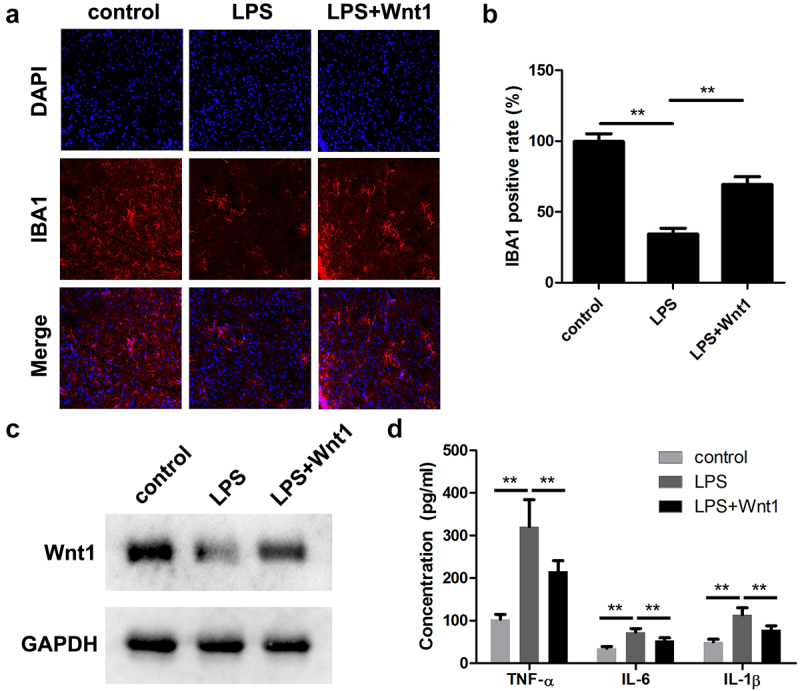
Intrauterine infection induces inflammation-mediated microglial activation and brain injury. This study aimed to explore the regulatory mechanism of Wnt family member 1 (Wnt1) in intrauterine infection-mediated microglial polarization. The cell counting kit-8 (CCK-8) assay was used to determine the viability of microglia, and cytokine expression levels were determined using enzyme linked immunosorbent assay (ELISA) kits and real-time quantitative PCR (RT-qPCR). The number of CD206 and CD16/32 cells was determined by flow cytometry. Wnt1 expression was analyzed using western blotting and immunofluorescence. Moreover, an assay was performed to verify the role of WNT1 in inflammation-sensitized brain injury in newborn mice. Lipopolysaccharide (LPS) exposure resulted in a decrease in microglial cell viability while increasing the expression levels of inflammatory cytokines (TNF-α, IL-6, and IL-1β), simultaneously promoting M1-type microglial conversion. However, these effects were rescued by overexpression of Wnt1, which was expressed less in microglia exposed to LPS and . Here, we found that Wnt1 activated the LKB1-AMPK pathway, and the inhibition of LKB1 attenuated the rescue effects of Wnt1. In addition, LPS exposure reduced the autophagy of microglia, and Wnt1 overexpression enhanced the autophagy, but this effect was reversed by treatment with an LKB1 inhibitor. Wnt1 activated LKB1 to suppress inflammation-mediated activation of microglia, promote M2-type microglia conversion via the AMPK pathway, and alleviate inflammation-sensitized neonatal brain injuries. This provides a potential avenue for the treatment of neonatal brain injuries.
宫内感染诱导炎症介导的小胶质细胞激活和脑损伤。本研究旨在探讨 Wnt 家族成员 1(Wnt1)在宫内感染介导的小胶质细胞极化中的调节机制。使用细胞计数试剂盒-8(CCK-8)测定小胶质细胞活力,酶联免疫吸附测定(ELISA)试剂盒和实时定量 PCR(RT-qPCR)测定细胞因子表达水平。通过流式细胞术测定 CD206 和 CD16/32 细胞的数量。使用 Western blot 和免疫荧光分析 Wnt1 表达。此外,进行了一项 以验证 WNT1 在新生小鼠炎症敏感脑损伤中的作用。脂多糖(LPS)暴露导致小胶质细胞活力降低,同时增加炎症细胞因子(TNF-α、IL-6 和 IL-1β)的表达水平,促进 M1 型小胶质细胞转化。然而,过表达 Wnt1 可挽救这些效应,LPS 暴露的小胶质细胞中 Wnt1 的表达减少。在这里,我们发现 Wnt1 激活了 LKB1-AMPK 通路,而 LKB1 的抑制减弱了 Wnt1 的挽救作用。此外,LPS 暴露降低了小胶质细胞的自噬,Wnt1 过表达增强了自噬,但 LKB1 抑制剂的处理逆转了这种效应。Wnt1 激活 LKB1 抑制炎症介导的小胶质细胞激活,通过 AMPK 通路促进 M2 型小胶质细胞转化,并减轻炎症敏感的新生儿脑损伤。这为新生儿脑损伤的治疗提供了一个潜在途径。