1 Department of Critical Care Medicine, The Fifth Peoples' Hospital of Dongguan, Dongguan Hospital Affiliated to Jinan University, Dongguan, China.
2 Department of Critical Care Medicine, General Hospital of Southern Theater Command, Key Laboratory of Tropical Trauma Care and Tissue Repair of PLA, Guangzhou, China.
Int J Immunopathol Pharmacol. 2019 Jan-Dec;33:2058738419828891. doi: 10.1177/2058738419828891.
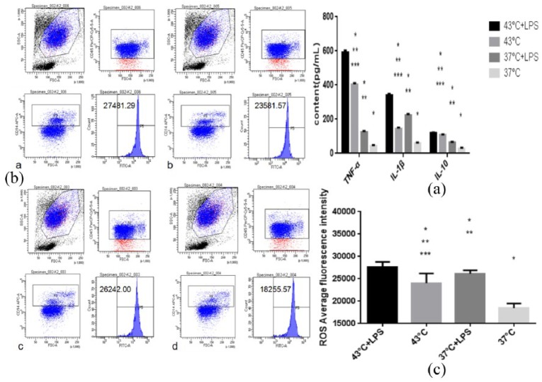
The purpose of this study was to focus on the underlying relationship between the hyperactivity for the peripheral monocytes and heat stroke by investigating the inflammatory oxidative activity of and the expression of superficial molecules. Peripheral blood samples were collected from 10 healthy adult volunteers. Human blood monocytes were isolated by density gradient centrifugation and sequent adherent culture. The objectives were divided into four groups: 43°C heat stress combined with lipopolysaccharide (LPS) group, 43°C heat stress group, LPS group, and control group. There were 10 cases in each group. An enzyme-linked immunosorbent assay (ELISA) test was used to measure the concentrations of supernatant inflammatory mediators (tumor necrosis factor alpha (TNF-α), interleukin-1β (IL-1β) and interleukin-10 (IL-10)). After loaded by 2,7-Dichlorodi-hydrofluorescein-diacetate (DCFHDA) fluorescent probe, intracellular reactive oxygen species (ROS) levels were determined by a flow cytometry. After fluorescent microspheres incubation, the phagocytosis of monocytes was observed under a fluorescent microscope. Respectively, the flow cytometry and Western blot were used to evaluate the level of triggering receptor expressed on myeloid cells-1 (TREM-1) and Toll-like receptor-4 (TLR-4) on the monocytes. Furthermore, the mRNA expression of TREM-1 and TLR-4 was detected by real-time polymerase chain reaction (RT-PCR). The heat stress combined with LPS stimulation promoted the peripheral monocytes to produce inflammatory mediators (TNF-α, IL-1β, and IL-10) and release ROS. Otherwise, such complex strike significantly suppressed the phagocytic activity of monocytes in peripheral blood. Moreover, the expression of TREM-1, TLR-4 and CD86 was measured by the flow cytometry on peripheral monocytes which were respectively promoted by the union of heat stress and LPS. The results of Western blot and RT-PCR demonstrated the similar kinetics on these superficial molecules (TREM-1, TLR-4, and CD86) stimulated by the combination of heat stress and LPS. The underlying mechanism of the dysfunction for the peripheral monocytes may be related to the abnormal expression of superficial molecules TREM-1, TLR-4, and CD86 on the monocytes induced by heat stress and LPS.
本研究旨在通过研究外周单核细胞活性与中暑之间的潜在关系,探讨炎症氧化活性和表面分子的表达。采集 10 名健康成年志愿者的外周血样本。通过密度梯度离心和连续贴壁培养分离人外周血单核细胞。将实验对象分为四组:43°C 热应激联合脂多糖(LPS)组、43°C 热应激组、LPS 组和对照组,每组 10 例。采用酶联免疫吸附试验(ELISA)检测上清液中炎症介质(肿瘤坏死因子-α(TNF-α)、白细胞介素-1β(IL-1β)和白细胞介素-10(IL-10))的浓度。用 2,7-二氯二氢荧光素二乙酸酯(DCFHDA)荧光探针加载后,通过流式细胞术测定细胞内活性氧(ROS)水平。经荧光微球孵育后,在荧光显微镜下观察单核细胞的吞噬作用。分别采用流式细胞术和 Western blot 检测单核细胞上髓样细胞触发受体-1(TREM-1)和 Toll 样受体-4(TLR-4)的表达水平,并通过实时聚合酶链反应(RT-PCR)检测 TREM-1 和 TLR-4 的 mRNA 表达。热应激联合 LPS 刺激促进外周血单核细胞产生炎症介质(TNF-α、IL-1β和 IL-10)并释放 ROS。此外,这种复合打击明显抑制外周血单核细胞的吞噬活性。此外,通过流式细胞术检测外周血单核细胞上 TREM-1、TLR-4 和 CD86 的表达,热应激和 LPS 的联合作用分别促进了这些表面分子的表达。Western blot 和 RT-PCR 的结果表明,热应激和 LPS 联合作用对这些表面分子(TREM-1、TLR-4 和 CD86)的刺激具有相似的动力学。外周单核细胞功能障碍的潜在机制可能与热应激和 LPS 诱导的单核细胞表面分子 TREM-1、TLR-4 和 CD86 的异常表达有关。