Department of Neurology, University of Tennessee Health Science Center, Memphis, Tennessee, United States of America.
Office of the Saskatchewan Multiple Sclerosis Clinical Research Chair, University of Saskatchewan, Saskatoon, Saskatchewan, Canada.
PLoS One. 2019 Feb 15;14(2):e0212357. doi: 10.1371/journal.pone.0212357. eCollection 2019.
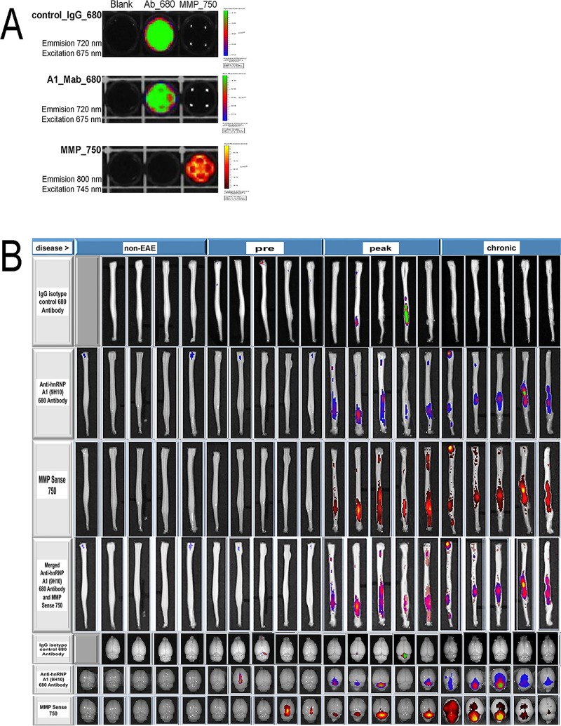
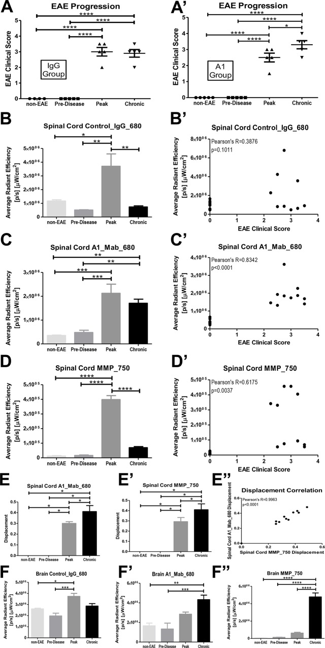
Antibodies, including antibodies to the RNA binding protein heterogeneous nuclear ribonucleoprotein A1, have been shown to contribute to the pathogenesis of multiple sclerosis, thus it is important to assess their biological activity using animal models of disease. Near-infrared optical imaging of fluorescently labeled antibodies and matrix metalloproteinase activity were measured and quantified in an animal model of multiple sclerosis, experimental autoimmune encephalomyelitis. We successfully labeled, imaged and quantified the fluorescence signal of antibodies that localized to the central nervous system of mice with experimental autoimmune encephalomyelitis. Fluorescently labeled anti-heterogeneous nuclear ribonucleoprotein A1 antibodies persisted in the central nervous system of mice with experimental autoimmune encephalomyelitis, colocalized with matrix metalloproteinase activity, correlated with clinical disease and shifted rostrally within the spinal cord, consistent with experimental autoimmune encephalomyelitis being an ascending paralysis. The fluorescent antibody signal also colocalized with matrix metalloproteinase activity in brain. Previous imaging studies in experimental autoimmune encephalomyelitis analyzed inflammatory markers such as cellular immune responses, dendritic cell activity, blood brain barrier integrity and myelination, but none assessed fluorescently labeled antibodies within the central nervous system. This data suggests a strong association between autoantibody localization and disease. This system can be used to detect other antibodies that might contribute to the pathogenesis of autoimmune diseases of the central nervous system including multiple sclerosis.
抗体,包括针对 RNA 结合蛋白异质性核核糖核蛋白 A1 的抗体,已被证明有助于多发性硬化症的发病机制,因此使用疾病的动物模型评估其生物学活性非常重要。在多发性硬化症的实验性自身免疫性脑脊髓炎动物模型中,测量和定量了荧光标记抗体和基质金属蛋白酶活性的近红外光学成像。我们成功地对实验性自身免疫性脑脊髓炎小鼠的中枢神经系统进行了荧光标记、成像和定量,荧光标记的抗异质性核核糖核蛋白 A1 抗体在实验性自身免疫性脑脊髓炎小鼠的中枢神经系统中持续存在,与基质金属蛋白酶活性共定位,与临床疾病相关,并在脊髓内向前迁移,与实验性自身免疫性脑脊髓炎是一种上升性瘫痪相一致。荧光抗体信号也与脑内的基质金属蛋白酶活性共定位。实验性自身免疫性脑脊髓炎的先前成像研究分析了细胞免疫反应、树突状细胞活性、血脑屏障完整性和髓鞘形成等炎症标志物,但没有评估中枢神经系统内的荧光标记抗体。这些数据表明自身抗体定位与疾病之间存在很强的关联。该系统可用于检测可能有助于中枢神经系统自身免疫性疾病(包括多发性硬化症)发病机制的其他抗体。