Australian Institute for Bioengineering and Nanotechnology (Building 75), The University of Queensland, Cooper Rd., St Lucia, Brisbane, QLD, 4072, Australia.
Laboratory of Immuno-Oncology, Department of Medical Oncology, Fujian Provincial Cancer Hospital &Institute, Fuzhou, 350014, China.
BMC Cancer. 2019 Feb 15;19(1):153. doi: 10.1186/s12885-019-5364-3.
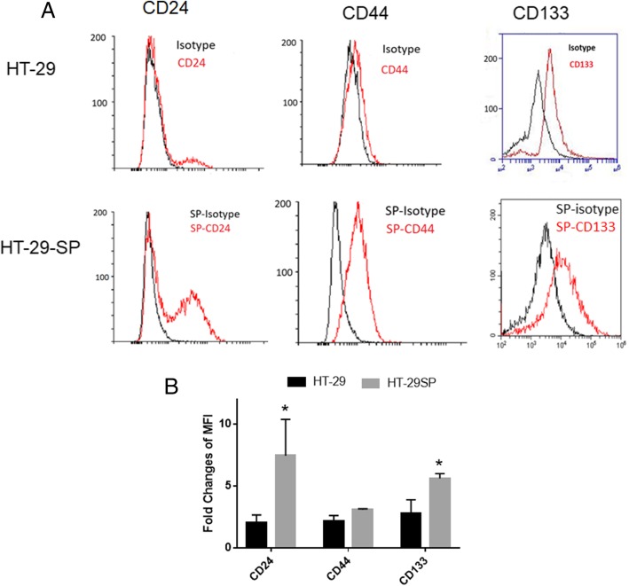
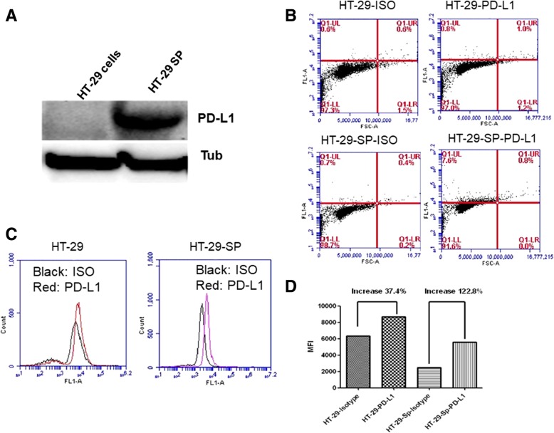
Programmed cell death ligand 1 (PD-L1) is an important immune-inhibitory protein expressed on cancer cells to mediate cancer escape through interaction with PD-1 expressed on activated T lymphocytes (T cells). Previously, we reported that colon and breast cancer stem cells (CSCs) expressed much higher levels of PD-L1 than their parental cells, suggesting they will be more resistant to immune attack.
We investigated the underlining mechanism of PD-L1 increase in colon CSCs, with a special focus on the effect of insulin and epithelial growth factor (EGF), the two fundamental components to sustain the metabolism and stemness in the culture of CSCs.
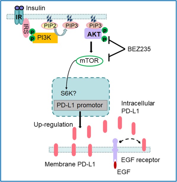
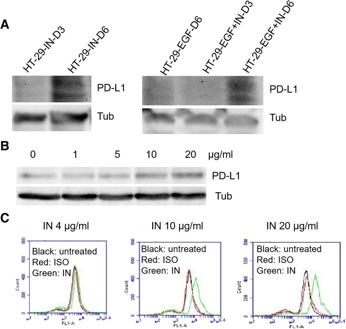
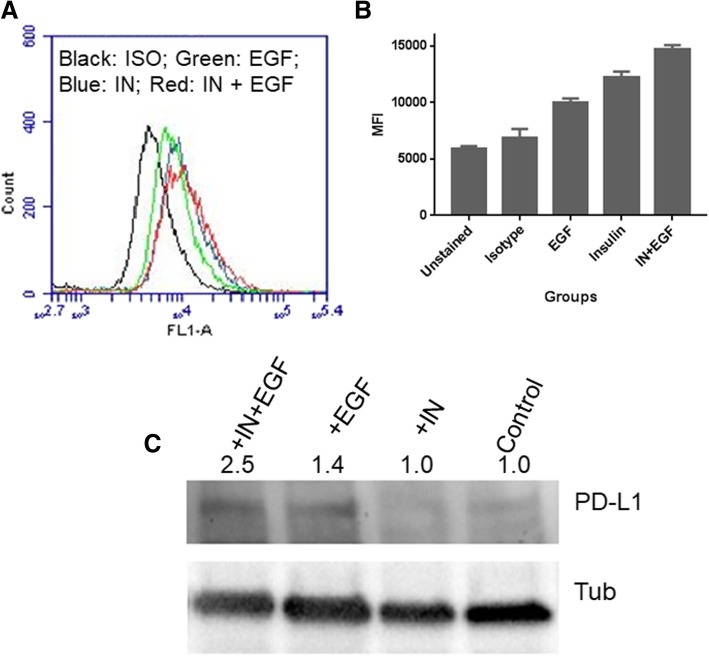
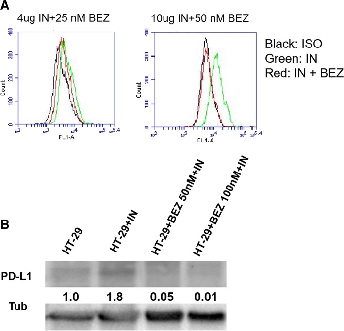
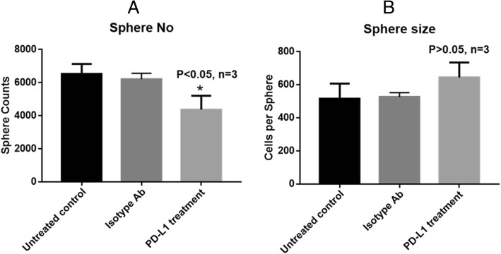
We found that insulin increased the total and surface PD-L1 levels through PI3K/Akt/mTOR pathway as the increase could be inhibited by the dual inhibitor of the pathway, BEZ235. EGF didn't affect the total PD-L1 levels of CSCs but increased the cell surface protein levels by flow cytometry analysis, indicating EGF promotes the transport of PD-L1 to the cell surface. Blocking cell surface PD-L1 with a specific antibody resulted in a significant reduction of tumour sphere formation but didn't interfere with the sphere growth, suggesting that cell surface PD-L1 may act as an adhering molecule for CSCs.
Apart from the essential roles in metabolism and stemness, insulin and EGF involve in up-regulation of PD-L1 expression in colon CSCs, therefore the inhibition of insulin and EGF/EGFR pathways can be considered for cancer immunotherapy or combined with PD-1/PD-L1 antibody-based cancer immunotherapy to eliminate CSCs.
程序性死亡配体 1(PD-L1)是一种在癌细胞上表达的重要免疫抑制蛋白,通过与激活的 T 淋巴细胞(T 细胞)上表达的 PD-1 相互作用,介导癌症逃逸。此前,我们报道结肠和乳腺癌干细胞(CSC)表达的 PD-L1 水平远高于其亲本细胞,这表明它们将更能抵抗免疫攻击。
我们研究了结肠 CSC 中 PD-L1 增加的潜在机制,特别关注胰岛素和表皮生长因子(EGF)的作用,这两种基本成分是维持 CSC 培养物代谢和干性所必需的。
我们发现胰岛素通过 PI3K/Akt/mTOR 通路增加总 PD-L1 和表面 PD-L1 水平,该通路的双重抑制剂 BEZ235 可抑制这种增加。EGF 不影响 CSC 的总 PD-L1 水平,但通过流式细胞术分析增加细胞表面蛋白水平,表明 EGF 促进 PD-L1 向细胞表面的转运。用特异性抗体阻断细胞表面 PD-L1 导致肿瘤球形成显著减少,但不干扰球的生长,这表明细胞表面 PD-L1 可能作为 CSC 的黏附分子。
除了在代谢和干性方面的重要作用外,胰岛素和 EGF 还参与结肠 CSC 中 PD-L1 的上调,因此,抑制胰岛素和 EGF/EGFR 通路可用于癌症免疫治疗,或与 PD-1/PD-L1 抗体联合进行癌症免疫治疗,以消除 CSC。