Priftakis Dimitrios, Rondogianni Phivi, Datseris Ioannis
Department of Nuclear Medicine and Positron Emission Tomography/Computed Tomography, General Hospital of Athens "Evangelismos," Athens, Greece.
World J Nucl Med. 2019 Jan-Mar;18(1):71-73. doi: 10.4103/wjnm.WJNM_15_18.
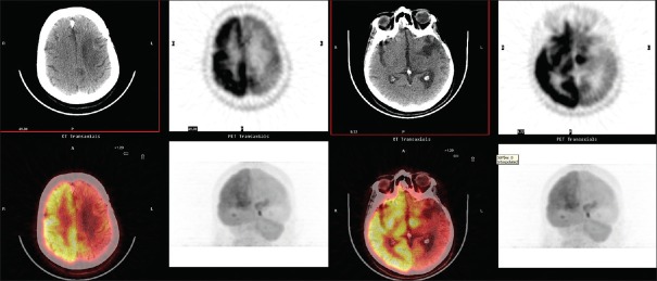
Crossed cerebellar diaschisis (CCD) represents the reduction of blood flow, metabolism, and oxygen consumption in the cerebellar hemisphere contralateral to a cerebral focal lesion. This phenomenon is the result of remote metabolic effects of cerebral lesions and it has been described since the first attempts for functional imaging of the brain, almost 40 years ago. Nevertheless, its clinical significance remains uncertain and new ways to use imaging of CCD for prognosis or assessment of novel therapies are being investigated. In this report, we present treatment for glioblastoma as a cause of CCD imaged on positron emission tomography/computed tomography with (F) fluoro-D-glucose in our department.
交叉性小脑失联络(CCD)表现为大脑局灶性病变对侧小脑半球血流、代谢及氧耗量减少。这种现象是脑部病变远程代谢效应的结果,自近40年前首次尝试进行脑功能成像以来就已被描述。然而,其临床意义仍不明确,目前正在研究利用CCD成像进行预后评估或新型疗法评估的新方法。在本报告中,我们介绍了在我们科室,以正电子发射断层扫描/计算机断层扫描(PET/CT)用(F)氟代-D-葡萄糖成像显示的胶质母细胞瘤作为CCD病因的治疗情况。