Division of Biochemical Toxicology, National Center for Toxicological Research, U.S. Food and Drug Administration, Jefferson, Arkansas, United States of America.
Center for Drug Evaluation and Research, U.S. Food and Drug Administration, Silver Spring, Maryland, United States of America.
PLoS One. 2019 Feb 19;14(2):e0210273. doi: 10.1371/journal.pone.0210273. eCollection 2019.
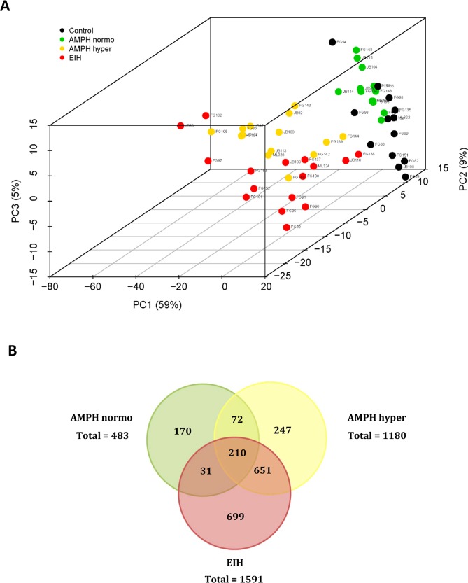
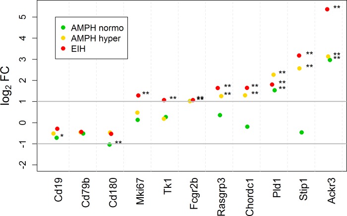
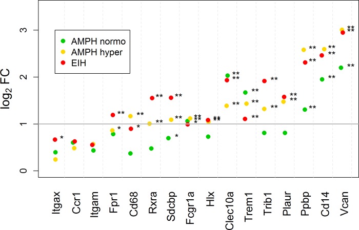
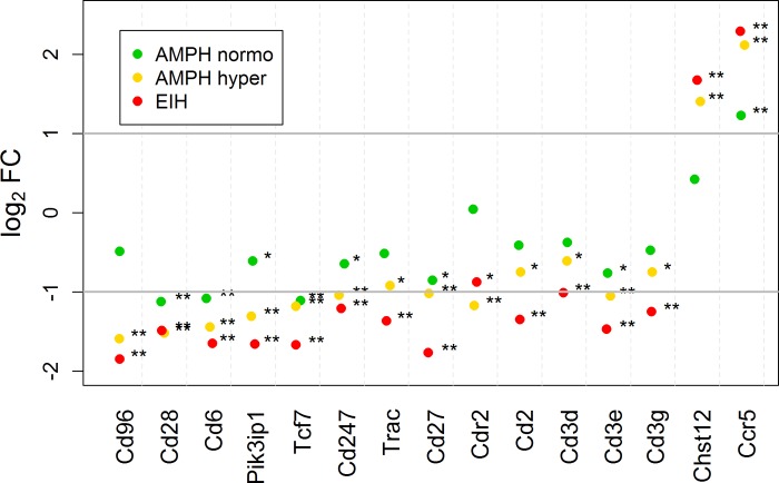
This work extends the understanding of how toxic exposures to amphetamine (AMPH) adversely affect the immune system and lead to tissue damage. Importantly, it determines which effects of AMPH are and are not due to pronounced hyperthermia. Whole blood messenger RNA (mRNA) and whole blood and serum microRNA (miRNA) transcripts were identified in adult male Sprague-Dawley rats after exposure to toxic AMPH under normothermic conditions, AMPH when it produces pronounced hyperthermia, or environmentally-induced hyperthermia (EIH). mRNA transcripts with large increases in fold-change in treated relative to control rats and very low expression in the control group were a rich source of organ-specific transcripts in blood. When severe hyperthermia was produced by either EIH or AMPH, significant increases in circulating organ-specific transcripts for liver (Alb, Fbg, F2), pancreas (Spink1), bronchi/lungs (F3, Cyp4b1), bone marrow (Np4, RatNP-3b), and kidney (Cesl1, Slc22a8) were observed. Liver damage was suggested also by increased miR-122 levels in the serum. Increases in muscle/heart-enriched transcripts were produced by AMPH even in the absence of hyperthermia. Expression increases in immune-related transcripts, particularly Cd14 and Vcan, indicate that AMPH can activate the innate immune system in the absence of hyperthermia. Most transcripts specific for T-cells decreased 50-70% after AMPH exposure or EIH, with the noted exception of Ccr5 and Chst12. This is probably due to T-cells leaving the circulation and down-regulation of these genes. Transcript changes specific for B-cells or B-lymphoblasts in the AMPH and EIH groups ranged widely from decreasing ≈ 40% (Cd19, Cd180) to increasing 30 to 100% (Tk1, Ahsa1) to increasing ≥500% (Stip1, Ackr3). The marked increases in Ccr2, Ccr5, Pld1, and Ackr3 produced by either AMPH or EIH observed in vivo provide further insight into the initial immune system alterations that result from methamphetamine and AMPH abuse and could modify risk for HIV and other viral infections.
这项工作扩展了我们对于安非他命(AMPH)的毒性暴露如何对免疫系统产生不利影响并导致组织损伤的认识。重要的是,它确定了 AMPH 的哪些作用是由于明显的体温升高引起的,哪些不是。在正常体温条件下,雄性 Sprague-Dawley 大鼠暴露于有毒的 AMPH 后,检测了其全血信使 RNA(mRNA)和全血及血清 microRNA(miRNA)转录本,以及在 AMPH 引起明显体温升高时,或在环境引起的体温升高(EIH)时。与对照组大鼠相比,处理组大鼠中折叠变化倍数大幅增加且对照组中表达水平极低的 mRNA 转录本是血液中器官特异性转录本的丰富来源。当 EIH 或 AMPH 产生严重的体温升高时,观察到循环中肝脏(Alb、Fbg、F2)、胰腺(Spink1)、支气管/肺(F3、Cyp4b1)、骨髓(Np4、RatNP-3b)和肾脏(Cesl1、Slc22a8)的器官特异性转录本显著增加。血清中 miR-122 水平的升高也提示存在肝损伤。即使在没有体温升高的情况下,AMPH 也会引起肌肉/心脏富集转录本的增加。免疫相关转录本,特别是 Cd14 和 Vcan 的表达增加,表明 AMPH 可以在没有体温升高的情况下激活先天免疫系统。大多数 T 细胞特异性转录本在 AMPH 暴露或 EIH 后降低了 50-70%,但 Ccr5 和 Chst12 除外。这可能是由于 T 细胞离开循环和这些基因的下调。AMPH 和 EIH 组中 B 细胞或 B 淋巴细胞特异性转录本的变化范围很广,从约 40%(Cd19、Cd180)减少到 30-100%(Tk1、Ahsa1)增加到 500%以上(Stip1、Ackr3)增加。无论是 AMPH 还是 EIH 引起的 Ccr2、Ccr5、Pld1 和 Ackr3 的明显增加,都进一步深入了解了源自安非他命和 AMPH 滥用的初始免疫系统改变,并可能改变 HIV 和其他病毒感染的风险。