Laboratory of Clinical Chemistry & Hematology, University Medical Center Utrecht, Utrecht University, Utrecht, The Netherlands.
Van Creveldkliniek, University Medical Center Utrecht, Utrecht University, Utrecht, The Netherlands.
Am J Hematol. 2019 May;94(5):575-584. doi: 10.1002/ajh.25443. Epub 2019 Mar 8.
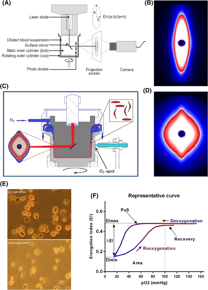
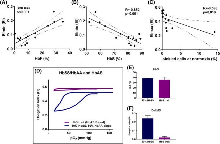
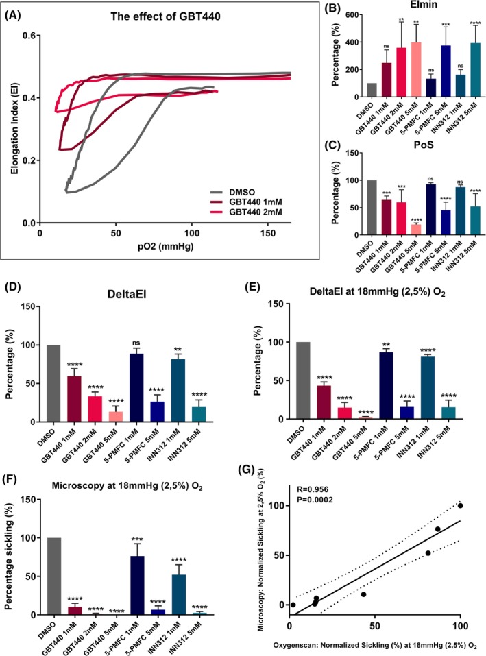
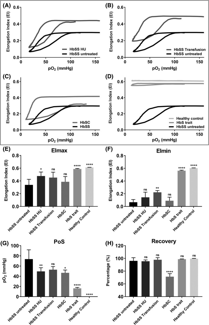
In sickle cell disease (SCD), sickle hemoglobin (HbS) polymerizes upon deoxygenation, resulting in sickling of red blood cells (RBCs). These sickled RBCs have strongly reduced deformability, leading to vaso-occlusive crises and chronic hemolytic anemia. To date, there are no reliable laboratory parameters or assays capable of predicting disease severity or monitoring treatment effects. We here report on the oxygenscan, a newly developed method to measure RBC deformability (expressed as Elongation Index - EI) as a function of pO . Upon a standardized, 22 minute, automated cycle of deoxygenation (pO median 16 mmHg ± 0.17) and reoxygenation, a number of clinically relevant parameters are produced in a highly reproducible manner (coefficients of variation <5%). In particular, physiological modulators of oxygen affinity, such as, pH and 2,3-diphosphoglycerate showed a significant correlation (respectively R = -0.993 and R = 0.980) with Point of Sickling (PoS ), which is defined as the pO where a 5% decrease in EI is observed during deoxygenation. Furthermore, in vitro treatment with antisickling agents, including GBT440, which alter the oxygen affinity of hemoglobin, caused a reproducible left-shift of the PoS, indicating improved deformability at lower oxygen tensions. When RBCs from 21 SCD patients were analyzed, we observed a significantly higher PoS in untreated homozygous SCD patients compared to treated patients and other genotypes. We conclude that the oxygenscan is a state-of-the-art technique that allows for rapid analysis of sickling behavior in SCD patients. The method is promising for personalized treatment, development of new treatment strategies and could have potential in prediction of complications.
在镰状细胞病(SCD)中,脱氧时镰状血红蛋白(HbS)聚合,导致红细胞(RBC)镰变。这些镰状 RBC 的变形能力大大降低,导致血管阻塞性危象和慢性溶血性贫血。迄今为止,还没有可靠的实验室参数或检测方法能够预测疾病严重程度或监测治疗效果。我们在此报告氧扫描,这是一种新开发的测量 RBC 变形性(表示为伸长指数-EI)作为 pO 的函数的方法。在标准化的 22 分钟自动脱氧(pO 中位数 16mmHg±0.17)和复氧循环中,以高度可重复的方式产生了许多与临床相关的参数(变异系数<5%)。特别是,氧亲和力的生理调节剂,如 pH 和 2,3-二磷酸甘油酸与镰变点(PoS)显示出显著的相关性(分别为 R=-0.993 和 R=0.980),PoS 定义为在脱氧过程中 EI 下降 5%时的 pO。此外,体外用包括 GBT440 在内的抗镰变剂处理可导致 PoS 重现性左移,表明在较低氧张力下变形能力提高。当分析 21 名 SCD 患者的 RBC 时,我们观察到未经治疗的纯合 SCD 患者的 PoS 明显高于经治疗的患者和其他基因型。我们得出结论,氧扫描是一种先进的技术,允许快速分析 SCD 患者的镰变行为。该方法有望用于个性化治疗、新治疗策略的开发,并可能具有预测并发症的潜力。