Department of Neurology, University of Colorado School of Medicine, Aurora, Colorado, United States.
Department of Ophthalmology, University of Colorado School of Medicine, Aurora, Colorado, United States.
Invest Ophthalmol Vis Sci. 2019 Feb 1;60(2):704-711. doi: 10.1167/iovs.18-25801.
While VZV DNA and antigen have been detected in acute and chronic VZV keratitis, it is unclear whether productive infection of corneal cells is ongoing or whether residual, noninfectious VZV antigens elicit inflammation. Herein, we examined VZV-infected primary human corneal epithelial cells (HCECs) and keratocytes (HKs) to elucidate the pathogenesis of VZV keratitis.
HCECs and HKs were mock- or VZV infected. Seven days later, cells were examined for morphology, proinflammatory cytokine and matrix metalloproteinase (MMP) release, ability to recruit peripheral blood mononuclear cells (PBMCs) and neutrophils, and MMP substrate cleavage.
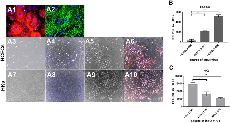
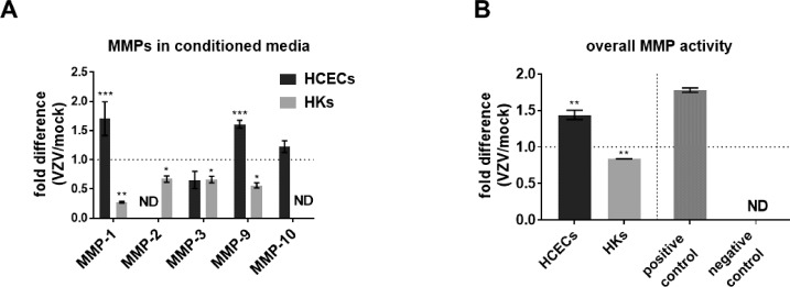
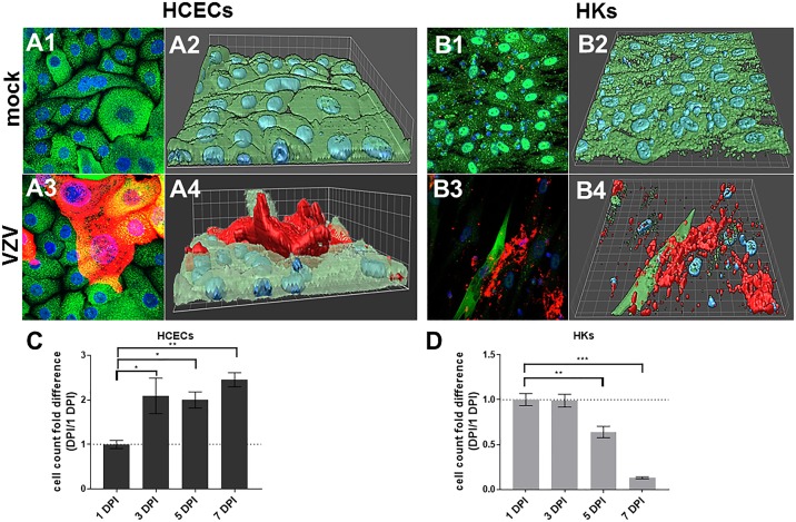
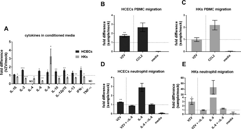
Both cell types synthesized infectious virus. VZV-infected HCECs proliferated, whereas VZV-infected HKs died. Compared to mock-infected cells, VZV-infected HCECs secreted significantly more IL-6, IL-8, IL-10, and IL-12p70 that were confirmed at the transcript level, and MMP-1 and MMP-9; conditioned supernatant attracted PBMCs and neutrophils and cleaved MMP substrates. In contrast, VZV-infected HKs suppressed cytokine secretion except for IL-8, which attracted neutrophils, and suppressed MMP release and substrate cleavage.
Overall, VZV-infected HCECs recapitulate findings of VZV keratitis with respect to epithelial cell proliferation, pseudodendrite formation and creation of a proinflammatory environment, providing an in vitro model for VZV infection of corneal epithelial cells. Furthermore, the proliferation and persistence of VZV-infected HCECs suggest that these cells may serve as viral reservoirs if immune clearance is incomplete. Finally, the finding that VZV-infected HKs die and suppress most proinflammatory cytokines and MMPs may explain the widespread death of these cells with unchecked viral spread due to ineffective recruitment of PBMCs.
虽然已在急性和慢性 VZV 角膜炎中检测到 VZV DNA 和抗原,但尚不清楚角膜细胞是否正在发生持续性感染,还是残留的非感染性 VZV 抗原引发炎症。在此,我们研究了 VZV 感染的原代人角膜上皮细胞(HCEC)和角膜基质细胞(HK),以阐明 VZV 角膜炎的发病机制。
HCEC 和 HK 被 mock 或 VZV 感染。7 天后,检查细胞形态、促炎细胞因子和基质金属蛋白酶(MMP)释放、募集外周血单核细胞(PBMC)和中性粒细胞的能力以及 MMP 底物裂解。
两种细胞类型均合成有感染性病毒。VZV 感染的 HCEC 增殖,而 VZV 感染的 HK 则死亡。与 mock 感染的细胞相比,VZV 感染的 HCEC 分泌了更多的 IL-6、IL-8、IL-10 和 IL-12p70,这些细胞因子在转录水平得到了证实,同时还分泌了 MMP-1 和 MMP-9;条件培养基吸引了 PBMC 和中性粒细胞,并裂解了 MMP 底物。相比之下,VZV 感染的 HK 抑制了细胞因子的分泌,除了 IL-8 之外,它还吸引了中性粒细胞,并抑制了 MMP 的释放和底物的裂解。
总体而言,VZV 感染的 HCEC 再现了 VZV 角膜炎的发现,包括上皮细胞增殖、假树突形成和炎症环境的产生,为 VZV 感染角膜上皮细胞提供了体外模型。此外,VZV 感染的 HCEC 的增殖和持续存在表明,如果免疫清除不完全,这些细胞可能成为病毒储存库。最后,VZV 感染的 HK 死亡并抑制大多数促炎细胞因子和 MMP 的发现可能解释了由于 PBMC 招募无效导致的这些细胞的广泛死亡和不受控制的病毒传播。