School of Medicine and Public Health, The University of Newcastle, Callaghan, NSW, Australia.
Hunter Medical Research Institute, New Lambton Heights, NSW, Australia.
PLoS One. 2019 Feb 21;14(2):e0212230. doi: 10.1371/journal.pone.0212230. eCollection 2019.
The prevalence of heart failure increases in the aging population and following myocardial infarction (MI), yet the extracellular matrix (ECM) remodeling underpinning the development of aging- and MI-associated cardiac fibrosis remains poorly understood. A link between inflammation and fibrosis in the heart has long been appreciated, but has mechanistically remained undefined. We investigated the expression of a novel protein, extracellular matrix protein 1 (ECM1) in the aging and infarcted heart.
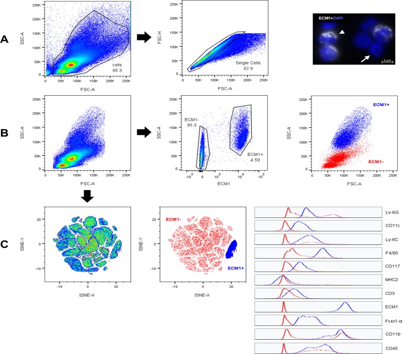
Young adult (3-month old) and aging (18-month old) C57BL/6 mice were assessed. Young mice were subjected to left anterior descending artery-ligation to induce MI, or transverse aortic constriction (TAC) surgery to induce pressure-overload cardiomyopathy. Left ventricle (LV) tissue was collected early and late post-MI/TAC. Bone marrow cells (BMCs) were isolated from young healthy mice, and subject to flow cytometry. Human cardiac fibroblast (CFb), myocyte, and coronary artery endothelial & smooth muscle cell lines were cultured; human CFbs were treated with recombinant ECM1. Primary mouse CFbs were cultured and treated with recombinant angiotensin-II or TGF-β1. Immunoblotting, qPCR and mRNA fluorescent in-situ hybridization (mRNA-FISH) were conducted on LV tissue and cells.
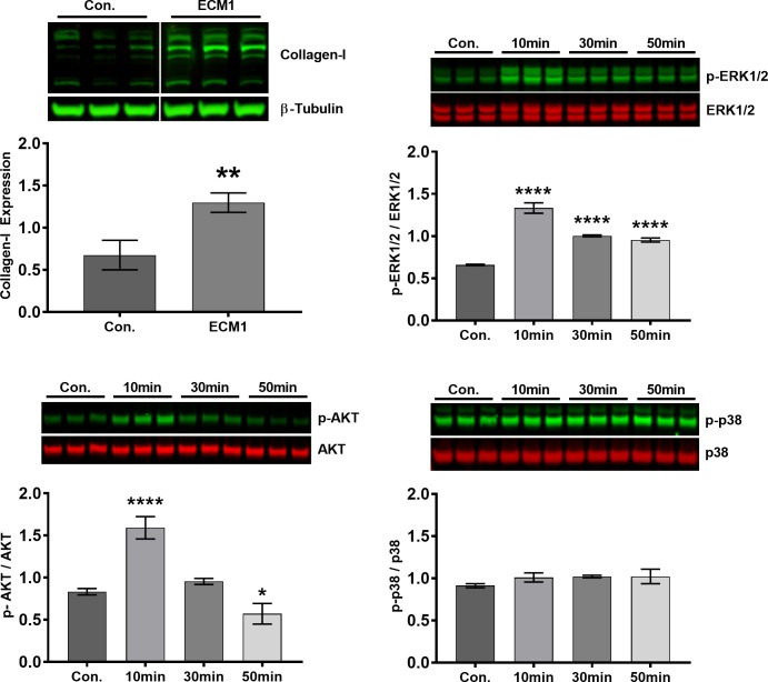
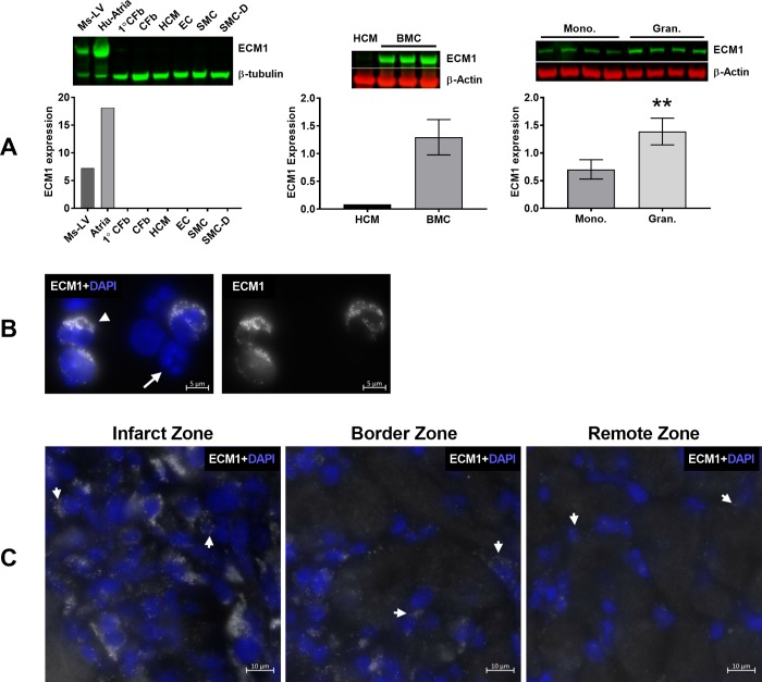
ECM1 expression was upregulated in the aging LV, and in the infarct zone of the LV early post-MI. No significant differences in ECM1 expression were found late post-MI or at any time-point post-TAC. ECM1 was not expressed in any resident cardiac cells, but ECM1 was highly expressed in BMCs, with high ECM1 expression in granulocytes. Flow cytometry of bone marrow revealed ECM1 expression in large granular leucocytes. mRNA-FISH revealed that ECM1 was indeed expressed by inflammatory cells in the infarct zone at day-3 post-MI. ECM1 stimulation of CFbs induced ERK1/2 and AKT activation and collagen-I expression, suggesting a pro-fibrotic role.
ECM1 expression is increased in ageing and infarcted hearts but is not expressed by resident cardiac cells. Instead it is expressed by bone marrow-derived granulocytes. ECM1 is sufficient to induce cardiac fibroblast stimulation in vitro. Our findings suggest ECM1 is released from infiltrating inflammatory cells, which leads to cardiac fibroblast stimulation and fibrosis in aging and MI. ECM1 may be a novel intermediary between inflammation and fibrosis.
心力衰竭的发病率随着人口老龄化和心肌梗死(MI)的发生而增加,但支持衰老和 MI 相关心肌纤维化发展的细胞外基质(ECM)重塑仍知之甚少。炎症与心脏纤维化之间的联系早已被人们所认识,但在机制上仍未得到明确界定。我们研究了一种新型蛋白,即细胞外基质蛋白 1(ECM1)在衰老和梗死心脏中的表达。
评估了年轻成年(3 个月大)和衰老(18 个月大)C57BL/6 小鼠。年轻小鼠接受左前降支结扎以诱导 MI,或接受升主动脉缩窄(TAC)手术以诱导压力超负荷性心肌病。MI/TAC 后早期和晚期收集左心室(LV)组织。从小鼠健康骨髓中分离出骨髓细胞(BMCs),并进行流式细胞术分析。培养人心房成纤维细胞(CFb)、心肌细胞和冠状动脉内皮和平滑肌细胞系;用重组 ECM1 处理人心房成纤维细胞。培养原代小鼠 CFb 并用重组血管紧张素-II 或 TGF-β1 处理。对 LV 组织和细胞进行免疫印迹、qPCR 和 mRNA 荧光原位杂交(mRNA-FISH)。
ECM1 在衰老的 LV 中以及 MI 后 LV 的梗死区表达上调。MI 后晚期或 TAC 后的任何时间点均未发现 ECM1 表达有显著差异。ECM1 不在任何常驻心肌细胞中表达,但在 BMC 中高度表达,在粒细胞中高表达。骨髓的流式细胞术显示 ECM1 在大颗粒白细胞中表达。mRNA-FISH 显示 ECM1 在 MI 后第 3 天的梗死区由炎症细胞表达。ECM1 刺激 CFb 诱导 ERK1/2 和 AKT 激活和胶原-I 表达,表明其具有促纤维化作用。
ECM1 在衰老和梗死的心脏中表达增加,但不在常驻心肌细胞中表达。相反,它由骨髓来源的粒细胞表达。ECM1 足以在体外诱导心脏成纤维细胞刺激。我们的研究结果表明,ECM1 是由浸润的炎症细胞释放的,导致衰老和 MI 中的心