Department of Radiology and Center for Imaging Science, Samsung Medical Center, Sungkyunkwan University School of Medicine, Seoul, Korea.
Department of Radiology, Chung-Ang University Hospital, Chung-Ang University College of Medicine, Seoul, Korea.
Korean J Radiol. 2019 Mar;20(3):513-521. doi: 10.3348/kjr.2018.0409.
To evaluate the efficacy of the morphologic-metabolic (M-M) dissociation sign based on computed tomography (CT) and fluorine-18-fluorodeoxyglucose positron emission tomography (PET)/CT in discriminating invasive mucinous adenocarcinoma (IMA) from invasive non-mucinous adenocarcinomas (ADCs) of the lung.
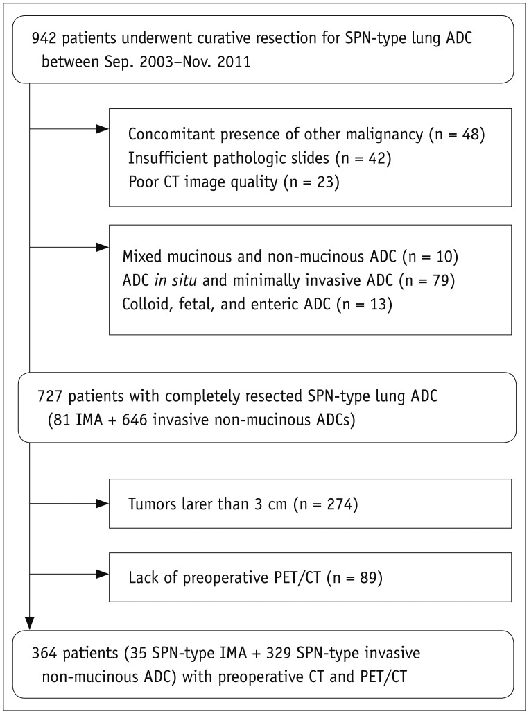
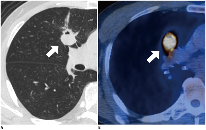
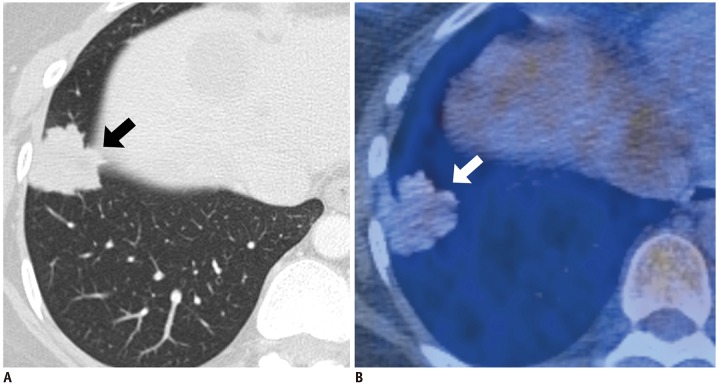
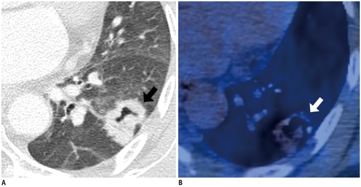
The Institutional Review Board approved this retrospective study. Among surgically resected solitary pulmonary nodule (SPN)-type ADCs (< 3 cm in diameter), 35 patients with IMAs and 329 with invasive non-mucinous ADCs were included. Morphologic malignancy was established if the tumor with lobulated or spiculated margin on CT presented a tumor shadow disappearance rate of < 0.5. The M-M dissociation sign was determined when a malignant-morphologic nodule on CT showed maximum standardized uptake value (SUVmax) < 3.5 on PET/CT.
Among 35 IMAs (size: 21 ± 7 mm, SUVmax: 1.8 ± 2.0) and 329 invasive non-mucinous ADCs (size: 21 ± 6 mm, SUVmax: 4.6 ± 4.2), the M-M dissociation sign was observed in 54% of IMAs (19/35) and 10% of invasive non-mucinous ADCs (34/329) ( < 0.001). The diagnostic performance of the sign in discriminating IMA from invasive non-mucinous ADCs showed a sensitivity of 54.3% (95% confidence interval [CI], 36.7-71.2), specificity 89.7% (95% CI, 85.9-92.7), positive predictive value 35.8% (95% CI, 26.5-46.5), and negative predictive value 94.9% (95% CI, 92.8-96.4). Multivariate analyses revealed metabolic benignity (odds ratio [OR] 2.99; 95% CI, 1.01-8.93; = 0.047) and M-M dissociation sign (OR 6.35; 95% CI, 2.76-14.62; < 0.001) to be significant predictors of SPN-type IMAs.
Identification of the absence of M-M dissociation sign is an accurate indicator for excluding IMA from SPN-type lung ADCs.
评估基于计算机断层扫描(CT)和氟-18-氟代脱氧葡萄糖正电子发射断层扫描(PET)/CT 的形态-代谢(M-M)分离征象在鉴别肺浸润性黏液腺癌(IMA)与浸润性非黏液性腺癌(ADC)中的疗效。
本回顾性研究经机构审查委员会批准。在手术切除的孤立性肺结节(SPN)型 ADC 中(直径<3cm),纳入 35 例 IMA 患者和 329 例浸润性非黏液性 ADC 患者。如果 CT 上呈分叶状或棘突状边缘的肿瘤呈现肿瘤阴影消失率<0.5,则确定形态恶性。当 CT 上的恶性形态结节在 PET/CT 上显示最大标准化摄取值(SUVmax)<3.5 时,确定 M-M 分离征象。
在 35 例 IMA(大小:21±7mm,SUVmax:1.8±2.0)和 329 例浸润性非黏液性 ADC 中(大小:21±6mm,SUVmax:4.6±4.2),54%的 IMA(19/35)和 10%的浸润性非黏液性 ADC(34/329)观察到 M-M 分离征象(<0.001)。该征象鉴别 IMA 与浸润性非黏液性 ADC 的诊断性能显示出 54.3%(95%置信区间[CI],36.7-71.2)的敏感性、89.7%(95%CI,85.9-92.7)的特异性、35.8%(95%CI,26.5-46.5)的阳性预测值和 94.9%(95%CI,92.8-96.4)的阴性预测值。多变量分析显示代谢良性(比值比[OR]2.99;95%CI,1.01-8.93;=0.047)和 M-M 分离征象(OR6.35;95%CI,2.76-14.62;<0.001)是 SPN 型 IMA 的显著预测因子。
识别不存在 M-M 分离征象是从 SPN 型肺 ADC 中排除 IMA 的准确指标。