Wang Tingting, Yang Yang, Liu Xinyue, Deng Jiajun, Wu Junqi, Hou Likun, Wu Chunyan, She Yunlang, Sun Xiwen, Xie Dong, Chen Chang
Department of Radiology, Shanghai Pulmonary Hospital, Tongji University School of Medicine, Shanghai, China.
Department of Thoracic Surgery, Shanghai Pulmonary Hospital, Tongji University School of Medicine, Shanghai, China.
Korean J Radiol. 2021 Apr;22(4):652-662. doi: 10.3348/kjr.2020.0454. Epub 2020 Nov 19.
To investigate the association between CT imaging features and survival outcomes in patients with primary invasive mucinous adenocarcinoma (IMA).
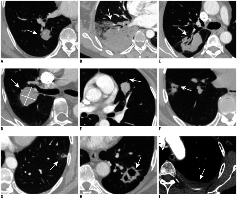
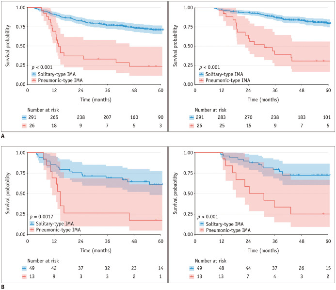
Preoperative CT image findings were consecutively evaluated in 317 patients with resected IMA from January 2011 to December 2015. The association between CT features and long-term survival were assessed by univariate analysis. The independent prognostic factors were identified by the multivariate Cox regression analyses. The survival comparison of IMA patients was investigated using the Kaplan-Meier method and propensity scores. Furthermore, the prognostic impact of CT features was assessed based on different imaging subtypes, and the results were adjusted using the Bonferroni method.
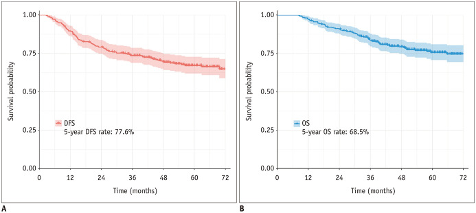
The median follow-up time was 52.8 months; the 5-year disease-free survival (DFS) and overall survival rates of resected IMAs were 68.5% and 77.6%, respectively. The univariate analyses of all IMA patients demonstrated that 15 CT imaging features, in addition to the clinicopathologic characteristics, significantly correlated with the recurrence or death of IMA patients. The multivariable analysis revealed that five of them, including imaging subtype ( = 0.002), spiculation ( < 0.001), tumor density ( = 0.008), air bronchogram ( < 0.001), emphysema ( < 0.001), and location ( = 0.029) were independent prognostic factors. The subgroup analysis demonstrated that pneumonic-type IMA had a significantly worse prognosis than solitary-type IMA. Moreover, for solitary-type IMAs, the most independent CT imaging biomarkers were air bronchogram and emphysema with an adjusted p value less than 0.05; for pneumonic-type IMA, the tumors with mixed consolidation and ground-glass opacity were associated with a longer DFS (adjusted = 0.012).
CT imaging features characteristic of IMA may provide prognostic information and individual risk assessment in addition to the recognized clinical predictors.
探讨原发性浸润性黏液腺癌(IMA)患者的CT影像特征与生存结局之间的关联。
对2011年1月至2015年12月期间317例接受IMA切除术患者的术前CT影像结果进行连续评估。通过单因素分析评估CT特征与长期生存之间的关联。采用多因素Cox回归分析确定独立预后因素。使用Kaplan-Meier法和倾向评分研究IMA患者的生存比较。此外,基于不同影像亚型评估CT特征的预后影响,并采用Bonferroni法对结果进行校正。
中位随访时间为52.8个月;切除IMA的5年无病生存率(DFS)和总生存率分别为68.5%和77.6%。对所有IMA患者的单因素分析表明,除临床病理特征外,15项CT影像特征与IMA患者的复发或死亡显著相关。多因素分析显示,其中5项,包括影像亚型(=0.002)、毛刺征(<0.001)、肿瘤密度(=0.008)、空气支气管征(<0.001)、肺气肿(<0.001)和位置(=0.029)是独立预后因素。亚组分析表明,肺炎型IMA的预后明显差于孤立型IMA。此外,对于孤立型IMA,最独立的CT影像生物标志物是空气支气管征和肺气肿,校正p值小于0.05;对于肺炎型IMA,具有混合实变和磨玻璃影的肿瘤与更长的DFS相关(校正=0.012)。
IMA的CT影像特征除了已认可的临床预测因素外,还可能提供预后信息和个体风险评估。