• 文献检索
  • 文档翻译
  • 深度研究
  • 学术资讯
  • Suppr Zotero 插件Zotero 插件
  • 邀请有礼
  • 套餐&价格
  • 历史记录
应用&插件
Suppr Zotero 插件Zotero 插件浏览器插件Mac 客户端Windows 客户端微信小程序
定价
高级版会员购买积分包购买API积分包
服务
文献检索文档翻译深度研究API 文档MCP 服务
关于我们
关于 Suppr公司介绍联系我们用户协议隐私条款
关注我们

Suppr 超能文献

核心技术专利:CN118964589B侵权必究
粤ICP备2023148730 号-1Suppr @ 2026

文献检索

告别复杂PubMed语法,用中文像聊天一样搜索,搜遍4000万医学文献。AI智能推荐,让科研检索更轻松。

立即免费搜索

文件翻译

保留排版,准确专业,支持PDF/Word/PPT等文件格式,支持 12+语言互译。

免费翻译文档

深度研究

AI帮你快速写综述,25分钟生成高质量综述,智能提取关键信息,辅助科研写作。

立即免费体验

运动感应超像素(MOSES)是一种系统的计算框架,用于量化和发现细胞运动表型。

Motion sensing superpixels (MOSES) is a systematic computational framework to quantify and discover cellular motion phenotypes.

机构信息

Ludwig Institute for Cancer Research, Nuffield Department of Clinical Medicine, University of Oxford, Oxford, United Kingdom.

Institute of Biomedical Engineering, Department of Engineering, University of Oxford, Oxford, United Kingdom.

出版信息

Elife. 2019 Feb 26;8:e40162. doi: 10.7554/eLife.40162.

DOI:10.7554/eLife.40162
PMID:30803483
原文链接:https://pmc.ncbi.nlm.nih.gov/articles/PMC6391079/
Abstract

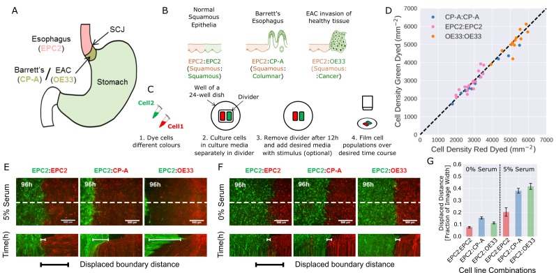
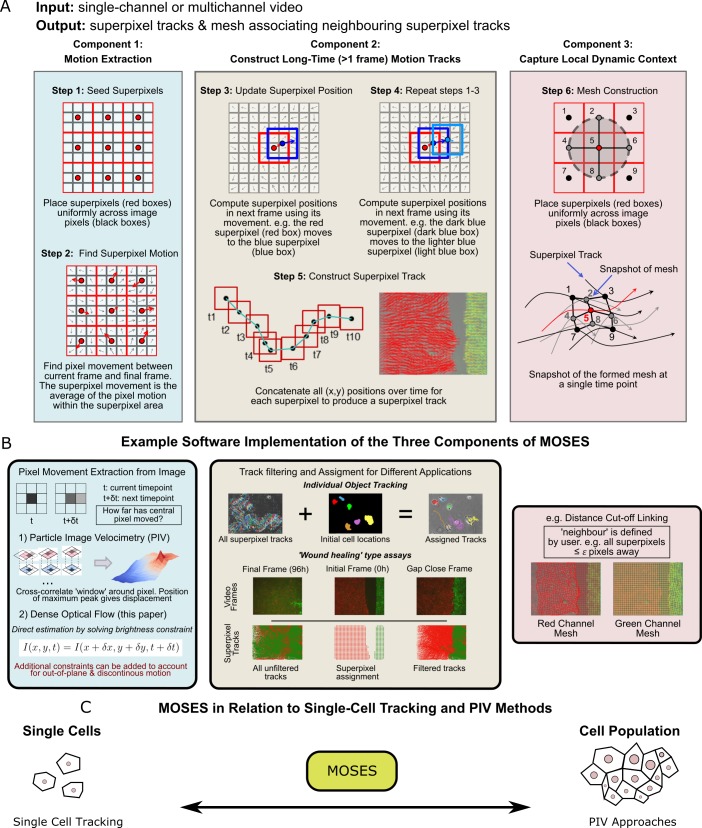
Correct cell/cell interactions and motion dynamics are fundamental in tissue homeostasis, and defects in these cellular processes cause diseases. Therefore, there is strong interest in identifying factors, including drug candidates that affect cell/cell interactions and motion dynamics. However, existing quantitative tools for systematically interrogating complex motion phenotypes in timelapse datasets are limited. We present Motion Sensing Superpixels (MOSES), a computational framework that measures and characterises biological motion with a unique superpixel 'mesh' formulation. Using published datasets, MOSES demonstrates single-cell tracking capability and more advanced population quantification than Particle Image Velocimetry approaches. From > 190 co-culture videos, MOSES motion-mapped the interactions between human esophageal squamous epithelial and columnar cells mimicking the esophageal squamous-columnar junction, a site where Barrett's esophagus and esophageal adenocarcinoma often arise clinically. MOSES is a powerful tool that will facilitate unbiased, systematic analysis of cellular dynamics from high-content time-lapse imaging screens with little prior knowledge and few assumptions.

摘要

正确的细胞/细胞相互作用和运动动力学是组织动态平衡的基础,这些细胞过程的缺陷会导致疾病。因此,人们强烈关注识别因素,包括影响细胞/细胞相互作用和运动动力学的药物候选物。然而,现有的用于系统研究时移数据集复杂运动表型的定量工具是有限的。我们提出了运动感应超像素(MOSES),这是一种计算框架,它使用独特的超像素“网格”公式来测量和描述生物运动。使用已发表的数据集,MOSES 展示了单细胞跟踪能力和比粒子图像测速方法更先进的群体量化能力。从 >190 个共培养视频中,MOSES 对人类食管鳞状上皮细胞和柱状细胞之间的相互作用进行了运动映射,模拟了食管鳞状柱状交界处,临床上 Barrett 食管和食管腺癌经常发生于此。MOSES 是一种强大的工具,它将促进从高内涵时移成像筛选中进行无偏、系统的细胞动力学分析,而无需先验知识和很少的假设。

https://cdn.ncbi.nlm.nih.gov/pmc/blobs/2f48/6391079/6be57c9e5019/elife-40162-fig5-figsupp2.jpg
https://cdn.ncbi.nlm.nih.gov/pmc/blobs/2f48/6391079/bd0d03f3a20e/elife-40162-fig1.jpg
https://cdn.ncbi.nlm.nih.gov/pmc/blobs/2f48/6391079/ec2f21bf8c1c/elife-40162-fig1-figsupp1.jpg
https://cdn.ncbi.nlm.nih.gov/pmc/blobs/2f48/6391079/e025e29a840d/elife-40162-fig1-figsupp2.jpg
https://cdn.ncbi.nlm.nih.gov/pmc/blobs/2f48/6391079/e9da5fd26e53/elife-40162-fig1-figsupp3.jpg
https://cdn.ncbi.nlm.nih.gov/pmc/blobs/2f48/6391079/6795b142141b/elife-40162-fig1-figsupp4.jpg
https://cdn.ncbi.nlm.nih.gov/pmc/blobs/2f48/6391079/31165b0b7945/elife-40162-fig1-figsupp5.jpg
https://cdn.ncbi.nlm.nih.gov/pmc/blobs/2f48/6391079/58517c627058/elife-40162-fig2.jpg
https://cdn.ncbi.nlm.nih.gov/pmc/blobs/2f48/6391079/d95378a2ff11/elife-40162-fig2-figsupp1.jpg
https://cdn.ncbi.nlm.nih.gov/pmc/blobs/2f48/6391079/17c52fe46ed8/elife-40162-fig2-figsupp2.jpg
https://cdn.ncbi.nlm.nih.gov/pmc/blobs/2f48/6391079/e9dd228ee2bc/elife-40162-fig3.jpg
https://cdn.ncbi.nlm.nih.gov/pmc/blobs/2f48/6391079/d65e9d127198/elife-40162-fig3-figsupp1.jpg
https://cdn.ncbi.nlm.nih.gov/pmc/blobs/2f48/6391079/1e0ca221c064/elife-40162-fig3-figsupp2.jpg
https://cdn.ncbi.nlm.nih.gov/pmc/blobs/2f48/6391079/065e48515ac1/elife-40162-fig3-figsupp3.jpg
https://cdn.ncbi.nlm.nih.gov/pmc/blobs/2f48/6391079/19347b2650d8/elife-40162-fig3-figsupp4.jpg
https://cdn.ncbi.nlm.nih.gov/pmc/blobs/2f48/6391079/a4c7ad05a3f0/elife-40162-fig3-figsupp5.jpg
https://cdn.ncbi.nlm.nih.gov/pmc/blobs/2f48/6391079/a443d9ff16fc/elife-40162-fig3-figsupp6.jpg
https://cdn.ncbi.nlm.nih.gov/pmc/blobs/2f48/6391079/d4db1877d653/elife-40162-fig3-figsupp7.jpg
https://cdn.ncbi.nlm.nih.gov/pmc/blobs/2f48/6391079/9fa112b6f98c/elife-40162-fig3-figsupp8.jpg
https://cdn.ncbi.nlm.nih.gov/pmc/blobs/2f48/6391079/db82cb2e39aa/elife-40162-fig3-figsupp9.jpg
https://cdn.ncbi.nlm.nih.gov/pmc/blobs/2f48/6391079/5bd56926da1e/elife-40162-fig3-figsupp10.jpg
https://cdn.ncbi.nlm.nih.gov/pmc/blobs/2f48/6391079/ac769e88e938/elife-40162-fig4.jpg
https://cdn.ncbi.nlm.nih.gov/pmc/blobs/2f48/6391079/f00dad0dbac4/elife-40162-fig4-figsupp1.jpg
https://cdn.ncbi.nlm.nih.gov/pmc/blobs/2f48/6391079/288908836bcb/elife-40162-fig4-figsupp2.jpg
https://cdn.ncbi.nlm.nih.gov/pmc/blobs/2f48/6391079/c97e71397b81/elife-40162-fig5.jpg
https://cdn.ncbi.nlm.nih.gov/pmc/blobs/2f48/6391079/acf74f4900a6/elife-40162-fig5-figsupp1.jpg
https://cdn.ncbi.nlm.nih.gov/pmc/blobs/2f48/6391079/6be57c9e5019/elife-40162-fig5-figsupp2.jpg

相似文献

1
Motion sensing superpixels (MOSES) is a systematic computational framework to quantify and discover cellular motion phenotypes.运动感应超像素(MOSES)是一种系统的计算框架,用于量化和发现细胞运动表型。
Elife. 2019 Feb 26;8:e40162. doi: 10.7554/eLife.40162.
2
Characterization of Biological Motion Using Motion Sensing Superpixels.使用运动感知超像素对生物运动进行表征
Bio Protoc. 2019 Sep 20;9(18):e3365. doi: 10.21769/BioProtoc.3365.
3
Correction: Motion sensing superpixels (MOSES) is a systematic computational framework to quantify and discover cellular motion phenotypes.更正:运动感知超像素(MOSES)是一个用于量化和发现细胞运动表型的系统性计算框架。
Elife. 2019 Jul 2;8:e49823. doi: 10.7554/eLife.49823.
4
Origins of Metaplasia in Barrett's Esophagus: Is this an Esophageal Stem or Progenitor Cell Disease?巴雷特食管化生的起源:这是一种食管干细胞或祖细胞疾病吗?
Dig Dis Sci. 2018 Aug;63(8):2005-2012. doi: 10.1007/s10620-018-5069-5.
5
Aberrant epithelial-mesenchymal Hedgehog signaling characterizes Barrett's metaplasia.异常的上皮-间充质 Hedgehog 信号特征表现为巴雷特食管化生。
Gastroenterology. 2010 May;138(5):1810-22. doi: 10.1053/j.gastro.2010.01.048. Epub 2010 Feb 4.
6
Inhibition of Notch signaling enhances transdifferentiation of the esophageal squamous epithelium towards a Barrett's-like metaplasia via KLF4.Notch信号通路的抑制通过KLF4增强食管鳞状上皮向巴雷特样化生的转分化。
Cell Cycle. 2014;13(24):3857-66. doi: 10.4161/15384101.2014.972875.
7
Forkhead box F1 induces columnar phenotype and epithelial-to-mesenchymal transition in esophageal squamous cells to initiate Barrett's like metaplasia.叉头框转录因子 F1 诱导食管鳞状细胞形成柱状表型和上皮间质转化,从而引发类似 Barrett 化生。
Lab Invest. 2021 Jun;101(6):745-759. doi: 10.1038/s41374-021-00534-4. Epub 2021 Jan 25.
8
Barrett's metaplasia develops from cellular reprograming of esophageal squamous epithelium due to gastroesophageal reflux.巴雷特化生是由于胃食管反流导致食管鳞状上皮细胞重编程而形成的。
Am J Physiol Gastrointest Liver Physiol. 2017 Jun 1;312(6):G615-G622. doi: 10.1152/ajpgi.00268.2016. Epub 2017 Mar 23.
9
Using cultured mammalian cells to study mitosis.利用培养的哺乳动物细胞研究有丝分裂。
Cold Spring Harb Protoc. 2012 Feb 1;2012(2):205-12. doi: 10.1101/pdb.ip067850.
10
Expression of the putative stem cell marker Musashi-1 in Barrett's esophagus and esophageal adenocarcinoma.Musashi-1 作为一种假定的干细胞标志物在 Barrett 食管和食管腺癌中的表达。
Dis Esophagus. 2010 Sep;23(7):580-9. doi: 10.1111/j.1442-2050.2010.01061.x. Epub 2010 Apr 29.

引用本文的文献

1
An integrated approach identifies the molecular underpinnings of murine anterior visceral endoderm migration.综合方法确定了小鼠前内脏内胚层迁移的分子基础。
Dev Cell. 2024 Sep 9;59(17):2347-2363.e9. doi: 10.1016/j.devcel.2024.05.014. Epub 2024 Jun 5.
2
Unsupervised Contour Tracking of Live Cells by Mechanical and Cycle Consistency Losses.基于机械和循环一致性损失的活细胞无监督轮廓跟踪
Proc IEEE Comput Soc Conf Comput Vis Pattern Recognit. 2023 Jun;2023:227-236. doi: 10.1109/cvpr52729.2023.00030. Epub 2023 Aug 22.
3
RhoU forms homo-oligomers to regulate cellular responses.

本文引用的文献

1
Long-lived force patterns and deformation waves at repulsive epithelial boundaries.排斥性上皮边界处的长寿命力模式和变形波。
Nat Mater. 2017 Oct;16(10):1029-1037. doi: 10.1038/nmat4972. Epub 2017 Sep 11.
2
Diverse roles of guanine nucleotide exchange factors in regulating collective cell migration.鸟嘌呤核苷酸交换因子在调节集体细胞迁移中的多种作用。
J Cell Biol. 2017 Jun 5;216(6):1543-1556. doi: 10.1083/jcb.201609095. Epub 2017 May 16.
3
Analysis of live cell images: Methods, tools and opportunities.活细胞图像分析:方法、工具与机遇。
RhoU 形成同型寡聚体以调节细胞反应。
J Cell Sci. 2024 Jan 15;137(2). doi: 10.1242/jcs.261645. Epub 2024 Jan 30.
4
Surface-guided computing to analyze subcellular morphology and membrane-associated signals in 3D.用于在三维空间中分析亚细胞形态和膜相关信号的表面引导计算。
bioRxiv. 2023 Apr 20:2023.04.12.536640. doi: 10.1101/2023.04.12.536640.
5
Surface-guided computing to analyze subcellular morphology and membrane-associated signals in 3D.用于分析三维亚细胞形态和膜相关信号的表面引导计算
ArXiv. 2023 Apr 12:arXiv:2304.06176v1.
6
Emerging machine learning approaches to phenotyping cellular motility and morphodynamics.新兴机器学习方法在细胞运动和形态动力学表型分析中的应用。
Phys Biol. 2021 Jun 17;18(4). doi: 10.1088/1478-3975/abffbe.
7
Characterization of Biological Motion Using Motion Sensing Superpixels.使用运动感知超像素对生物运动进行表征
Bio Protoc. 2019 Sep 20;9(18):e3365. doi: 10.21769/BioProtoc.3365.
8
DeepScratch: Single-cell based topological metrics of scratch wound assays.DeepScratch:基于单细胞的划痕实验拓扑指标
Comput Struct Biotechnol J. 2020 Aug 29;18:2501-2509. doi: 10.1016/j.csbj.2020.08.018. eCollection 2020.
9
Visualization of T Cell Migration in the Spleen Reveals a Network of Perivascular Pathways that Guide Entry into T Zones.脾脏中 T 细胞迁移的可视化显示了一个血管周途径网络,该网络指导 T 区进入。
Immunity. 2020 May 19;52(5):794-807.e7. doi: 10.1016/j.immuni.2020.03.010. Epub 2020 Apr 15.
10
Characterization of the Dynamic Behavior of Neutrophils Following Influenza Vaccination.流感疫苗接种后中性粒细胞动态行为的特征。
Front Immunol. 2019 Nov 20;10:2621. doi: 10.3389/fimmu.2019.02621. eCollection 2019.
Methods. 2017 Feb 15;115:65-79. doi: 10.1016/j.ymeth.2017.02.007. Epub 2017 Feb 27.
4
Endocytic reawakening of motility in jammed epithelia.在细胞外基质中被阻断的细胞重新获得运动能力。
Nat Mater. 2017 May;16(5):587-596. doi: 10.1038/nmat4848. Epub 2017 Jan 30.
5
TrackMate: An open and extensible platform for single-particle tracking.TrackMate:一个用于单粒子追踪的开放且可扩展的平台。
Methods. 2017 Feb 15;115:80-90. doi: 10.1016/j.ymeth.2016.09.016. Epub 2016 Oct 3.
6
Cell Painting, a high-content image-based assay for morphological profiling using multiplexed fluorescent dyes.细胞染色法,一种基于高内涵成像的多重荧光染料用于形态分析的方法。
Nat Protoc. 2016 Sep;11(9):1757-74. doi: 10.1038/nprot.2016.105. Epub 2016 Aug 25.
7
Seeds of Locally Aligned Motion and Stress Coordinate a Collective Cell Migration.局部对齐运动和应力的种子协调集体细胞迁移。
Biophys J. 2015 Dec 15;109(12):2492-2500. doi: 10.1016/j.bpj.2015.11.001.
8
Microscopy-Based High-Content Screening.基于显微镜的高通量筛选。
Cell. 2015 Dec 3;163(6):1314-25. doi: 10.1016/j.cell.2015.11.007.
9
Unjamming and cell shape in the asthmatic airway epithelium.哮喘气道上皮中的解阻塞与细胞形态
Nat Mater. 2015 Oct;14(10):1040-8. doi: 10.1038/nmat4357. Epub 2015 Aug 3.
10
Wound repair: role of immune-epithelial interactions.伤口修复:免疫-上皮相互作用的作用
Mucosal Immunol. 2015 Sep;8(5):959-68. doi: 10.1038/mi.2015.63. Epub 2015 Jul 15.