Department of Biomedical Engineering, Worcester Polytechnic Institute, Worcester, MA 01609, United States of America.
Vascular Biology Program and Department of Surgery, Boston Children's Hospital, Harvard Medical School, Boston, MA 02115, United States of America.
Phys Biol. 2021 Jun 17;18(4). doi: 10.1088/1478-3975/abffbe.
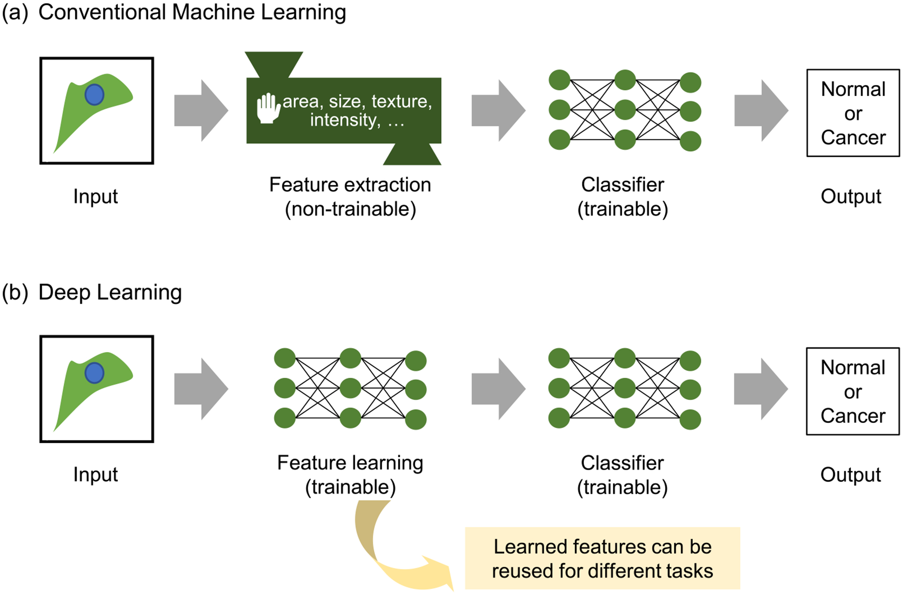
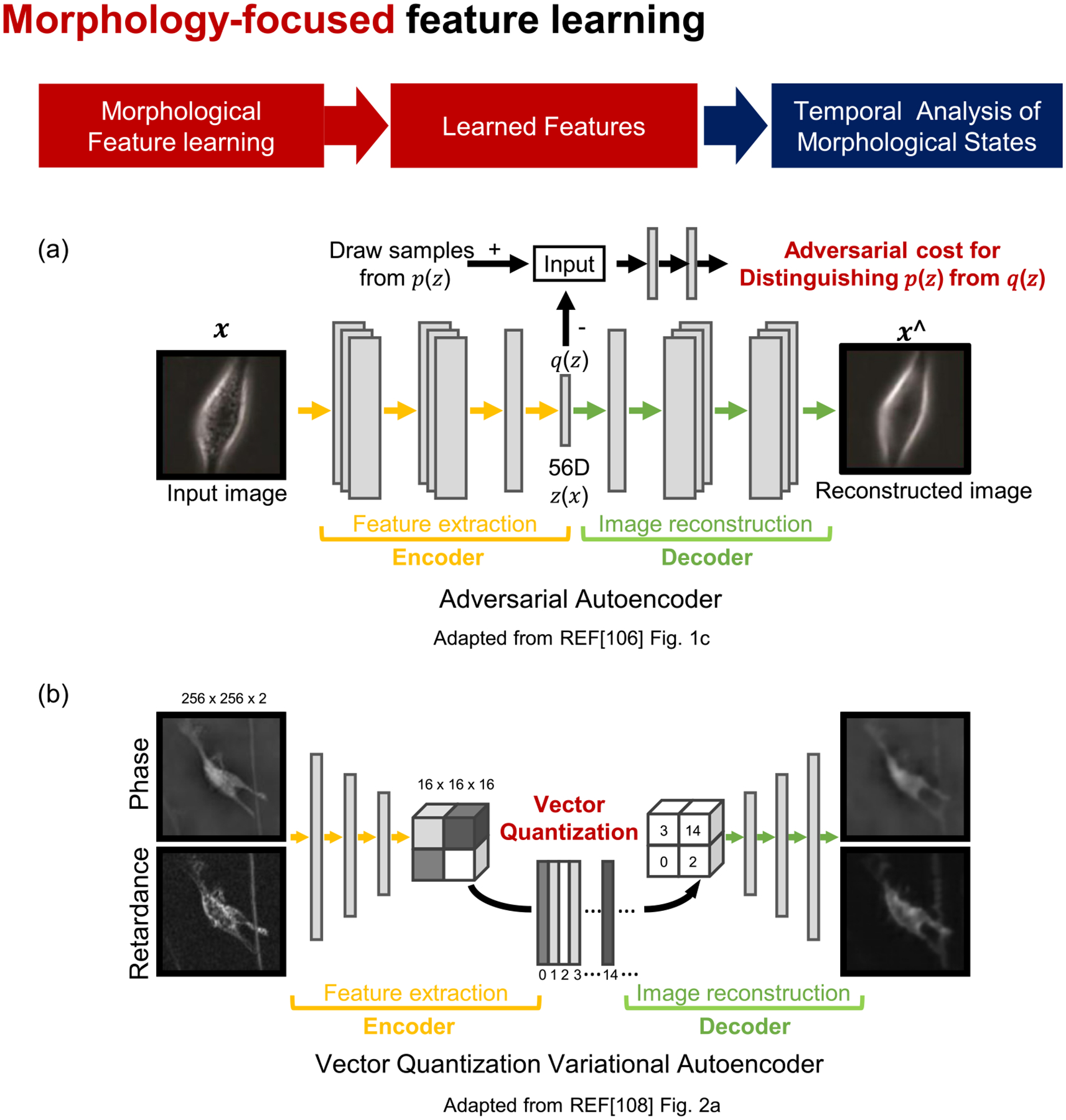
Cells respond heterogeneously to molecular and environmental perturbations. Phenotypic heterogeneity, wherein multiple phenotypes coexist in the same conditions, presents challenges when interpreting the observed heterogeneity. Advances in live cell microscopy allow researchers to acquire an unprecedented amount of live cell image data at high spatiotemporal resolutions. Phenotyping cellular dynamics, however, is a nontrivial task and requires machine learning (ML) approaches to discern phenotypic heterogeneity from live cell images. In recent years, ML has proven instrumental in biomedical research, allowing scientists to implement sophisticated computation in which computers learn and effectively perform specific analyses with minimal human instruction or intervention. In this review, we discuss how ML has been recently employed in the study of cell motility and morphodynamics to identify phenotypes from computer vision analysis. We focus on new approaches to extract and learn meaningful spatiotemporal features from complex live cell images for cellular and subcellular phenotyping.
细胞对分子和环境的干扰会产生不均匀的反应。表型异质性,即在相同条件下多种表型共存,给解释观察到的异质性带来了挑战。活细胞显微镜技术的进步使得研究人员能够以高时空分辨率获取前所未有的大量活细胞图像数据。然而,对细胞动力学进行表型分析是一项艰巨的任务,需要机器学习 (ML) 方法来从活细胞图像中识别表型异质性。近年来,ML 在生物医学研究中已被证明是非常有效的,它使科学家能够实施复杂的计算,让计算机在最小的人工指导或干预下学习并有效地执行特定的分析。在这篇综述中,我们讨论了 ML 最近如何被用于研究细胞迁移和形态动力学,以从计算机视觉分析中识别表型。我们专注于从复杂的活细胞图像中提取和学习有意义的时空特征的新方法,用于细胞和亚细胞表型分析。