Casey Eye Institute, Oregon Health & Science University, Portland, Oregon, United States.
Invest Ophthalmol Vis Sci. 2019 Feb 1;60(2):843-851. doi: 10.1167/iovs.18-26055.
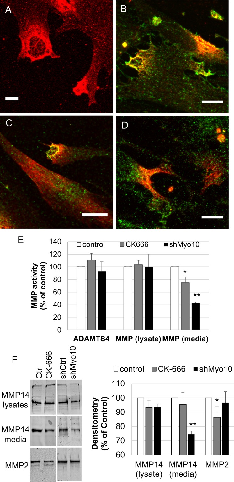
The actin cytoskeleton plays a key role in outflow regulation through the trabecular meshwork (TM). Although actin stress fibers are a target of glaucoma therapies, the role of other actin cellular structures is unclear. Myosin-X (Myo10) is an actin-binding protein that is involved in tunneling nanotube (TNT) and filopodia formation. Here, we inhibited Myo10 pharmacologically or by gene silencing to investigate the role of filopodia/TNTs in the TM.
Short hairpin RNA interference (RNAi) silencing lentivirus targeting myosin-X (shMyo10) was generated. Human anterior segments were perfused with shMyo10 or CK-666, an Arp2/3 inhibitor. Confocal microscopy investigated the colocalization of Myo10 with matrix metalloproteinase (MMPs). Western immunoblotting investigated the protein levels of MMPs and extracellular matrix (ECM) proteins. MMP activity and phagocytosis assays were performed.
CK-666 and shMyo10-silencing lentivirus caused a significant reduction in outflow rates in anterior segment perfusion culture, an ex vivo method to study intraocular pressure regulation. In human TM cells, Myo10 colocalized with MMP2, MMP14, and cortactin in podosome-like structures, which function as regions of focal ECM degradation. Furthermore, MMP activity, thrombospondin-1 and SPARC protein levels were significantly reduced in the media of CK-666-treated and shMyo10-silenced TM cells. However, neither Myo10 silencing or CK-666 treatment significantly affected phagocytic uptake.
Inhibiting filopodia/TNTs caused opposite effects on outflow compared with inhibiting stress fibers. Moreover, Myo10 may also play a role in focal ECM degradation in TM cells. Our results provide additional insight into the function of actin supramolecular assemblies and actin-binding proteins in outflow regulation.
细胞骨架肌动蛋白在小梁网(TM)流出调节中起关键作用。尽管肌动球蛋白应力纤维是青光眼治疗的靶点,但其他肌动蛋白细胞结构的作用尚不清楚。肌球蛋白-X(Myo10)是一种肌动蛋白结合蛋白,参与形成隧道纳米管(TNT)和丝状伪足。在这里,我们通过药理学或基因沉默抑制 Myo10,以研究 TM 中的丝状伪足/TNTs 的作用。
生成针对肌球蛋白-X(shMyo10)的短发夹 RNA 干扰(RNAi)沉默慢病毒。用 shMyo10 或 Arp2/3 抑制剂 CK-666 灌流人眼前段。共聚焦显微镜研究 Myo10 与基质金属蛋白酶(MMPs)的共定位。Western 免疫印迹法研究 MMPs 和细胞外基质(ECM)蛋白的蛋白水平。进行 MMP 活性和吞噬作用测定。
CK-666 和 shMyo10 沉默慢病毒导致前节灌注培养中流出率显著降低,这是一种研究眼压调节的离体方法。在人 TM 细胞中,Myo10 与 podosome 样结构中的 MMP2、MMP14 和 cortactin 共定位,后者作为局部 ECM 降解的区域起作用。此外,CK-666 处理和 shMyo10 沉默 TM 细胞培养基中的 MMP 活性、血栓反应蛋白-1 和 SPARC 蛋白水平显著降低。然而,Myo10 沉默或 CK-666 处理均未显著影响吞噬作用。
与抑制应力纤维相比,抑制丝状伪足/TNTs 对流出有相反的影响。此外,Myo10 也可能在 TM 细胞中的局部 ECM 降解中发挥作用。我们的结果为肌动蛋白超分子组装和肌动蛋白结合蛋白在流出调节中的作用提供了更多的见解。