Vranka Janice A, Kelley Mary J, Acott Ted S, Keller Kate E
Casey Eye Institute, Oregon Health & Science University, Portland, OR 97239, USA.
Casey Eye Institute, Oregon Health & Science University, Portland, OR 97239, USA.
Exp Eye Res. 2015 Apr;133:112-25. doi: 10.1016/j.exer.2014.07.014.
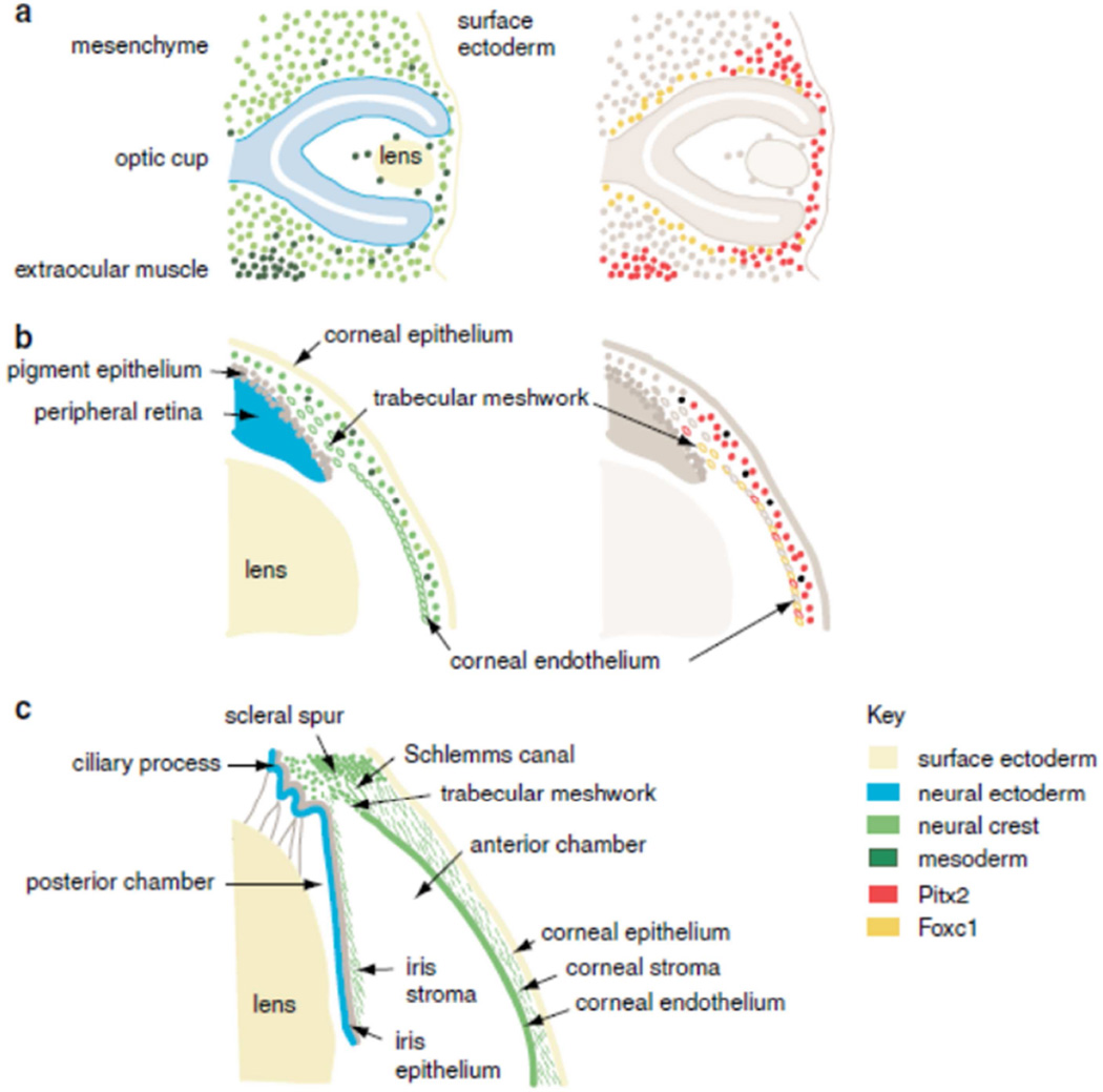
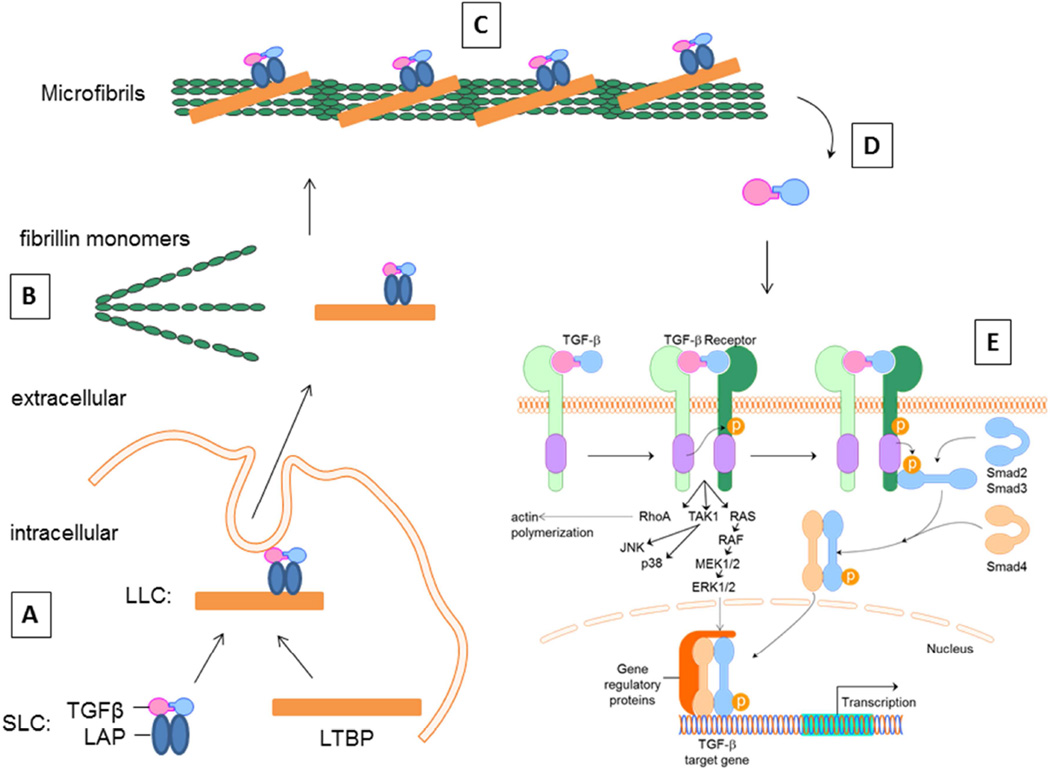
The trabecular meshwork (TM) is located in the anterior segment of the eye and is responsible for regulating the outflow of aqueous humor. Increased resistance to aqueous outflow causes intraocular pressure to increase, which is the primary risk factor for glaucoma. TM cells reside on a series of fenestrated beams and sheets through which the aqueous humor flows to exit the anterior chamber via Schlemm's canal. The outer trabecular cells are phagocytic and are thought to function as a pre-filter. However, most of the outflow resistance is thought to be from the extracellular matrix (ECM) of the juxtacanalicular region, the deepest portion of the TM, and from the inner wall basement membrane of Schlemm's canal. It is becoming increasingly evident that the extracellular milieu is important in maintaining the integrity of the TM. In glaucoma, not only have ultrastructural changes been observed in the ECM of the TM, and a significant number of mutations in ECM genes been noted, but the stiffness of glaucomatous TM appears to be greater than that of normal tissue. Additionally, TGFβ2 has been found to be elevated in the aqueous humor of glaucoma patients and is assumed to be involved in ECM changes deep with the juxtacanalicular region of the TM. This review summarizes the current literature on trabecular ECM as well as the development and function of the TM. Animal models and organ culture models targeting specific ECM molecules to investigate the mechanisms of glaucoma are described. Finally, the growing number of mutations that have been identified in ECM genes and genes that modulate ECM in humans with glaucoma are documented.
小梁网(TM)位于眼球前段,负责调节房水流出。房水流出阻力增加会导致眼压升高,这是青光眼的主要危险因素。TM细胞位于一系列有孔的梁和片上,房水通过这些结构经施莱姆管流出前房。小梁外层细胞具有吞噬作用,被认为起到预滤器的作用。然而,大部分流出阻力被认为来自近管区(TM最深的部分)的细胞外基质(ECM)以及施莱姆管的内壁基底膜。越来越明显的是,细胞外环境对于维持TM的完整性很重要。在青光眼中,不仅在TM的ECM中观察到超微结构变化,并且注意到ECM基因存在大量突变,而且青光眼TM的硬度似乎大于正常组织。此外,已发现青光眼患者房水中转化生长因子β2(TGFβ2)升高,并假定其参与TM近管区深处的ECM变化。本综述总结了关于小梁ECM以及TM的发育和功能的当前文献。描述了针对特定ECM分子以研究青光眼机制的动物模型和器官培养模型。最后,记录了在青光眼患者中已鉴定出的ECM基因和调节ECM的基因中越来越多的突变。