Department of Neuroscience, Cell Biology and Physiology, Wright State University Boonshoft School of Medicine, Dayton, Ohio, 45435, USA.
Department of Reproductive Medicine, University of California-San Diego, San Diego, California, 92093, USA.
Sci Rep. 2019 Feb 26;9(1):2742. doi: 10.1038/s41598-019-39426-5.
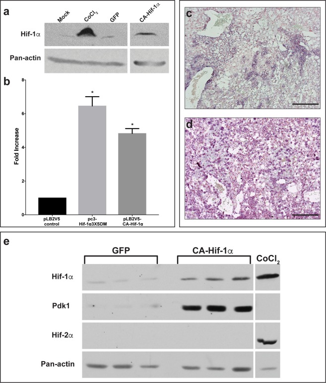
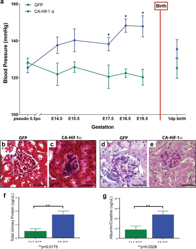
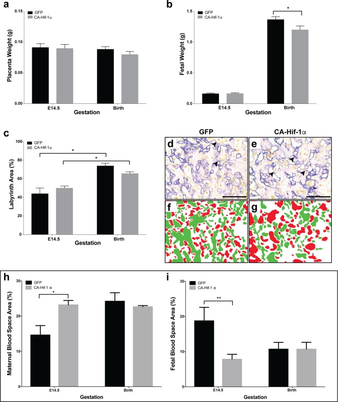
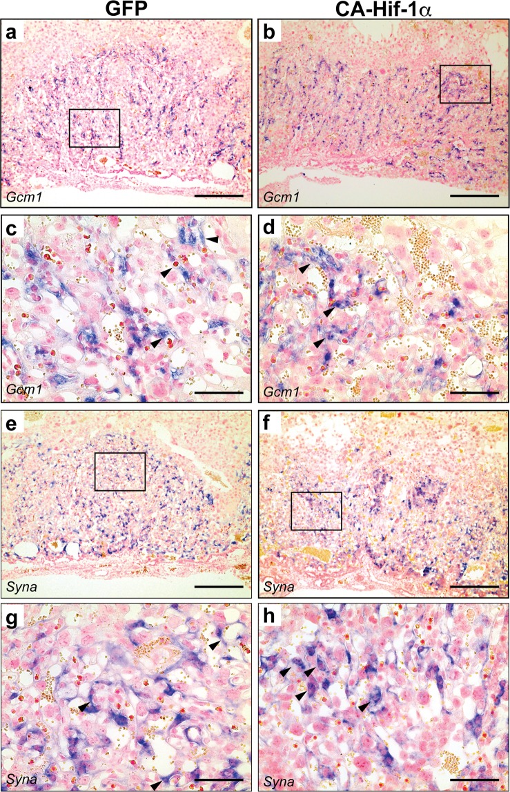
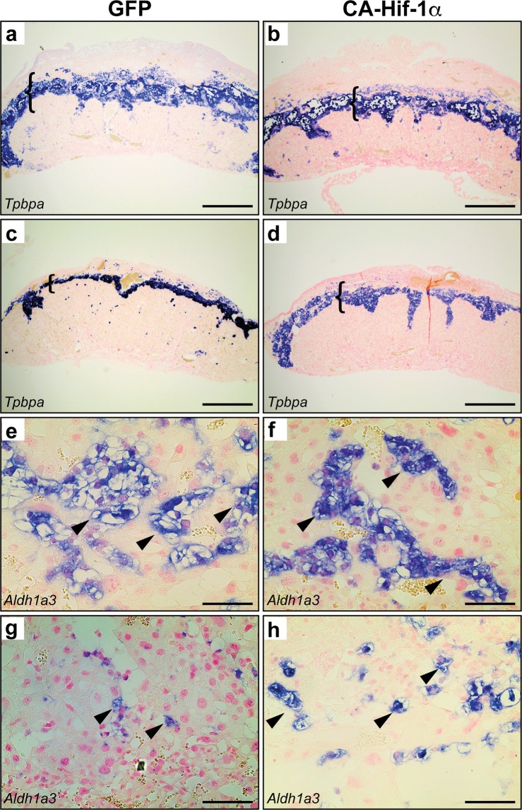
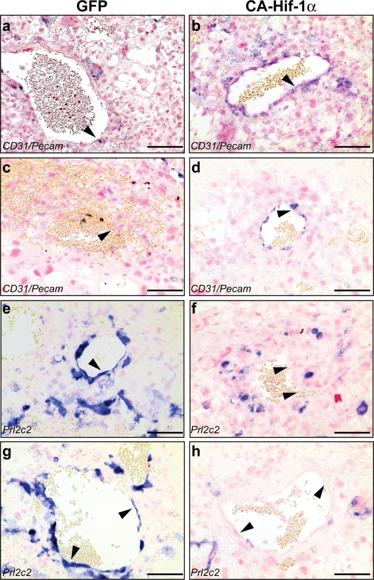
The placenta is an essential organ that is formed during pregnancy and its proper development is critical for embryonic survival. While several animal models have been shown to exhibit some of the pathological effects present in human preeclampsia, these models often do not represent the physiological aspects that have been identified. Hypoxia-inducible factor 1 alpha (Hif-1α) is a necessary component of the cellular oxygen-sensing machinery and has been implicated as a major regulator of trophoblast differentiation. Elevated levels of Hif-1α in the human placenta have been linked to the development of pregnancy-associated disorders, such as preeclampsia and fetal growth restriction. As oxygen regulation is a critical determinant for placentogenesis, we determined the effects of constitutively active Hif-1α, specifically in trophoblasts, on mouse placental development in vivo. Our research indicates that prolonged expression of trophoblast-specific Hif-1α leads to a significant decrease in fetal birth weight. In addition, we noted significant physiological alterations in placental differentiation that included reduced branching morphogenesis, alterations in maternal and fetal blood spaces, and failure to remodel the maternal spiral arteries. These placental alterations resulted in subsequent maternal hypertension with parturitional resolution and maternal kidney glomeruloendotheliosis with accompanying proteinuria, classic hallmarks of preeclampsia. Our findings identify Hif-1α as a critical molecular mediator of placental development and indicate that prolonged expression of Hif-1α, explicitly in placental trophoblasts causes maternal pathology and establishes a mouse model that significantly recapitulates the physiological and pathophysiological characteristics of preeclampsia with fetal growth restriction.
胎盘是一种在妊娠期间形成的重要器官,其正常发育对于胚胎的存活至关重要。虽然已经有几种动物模型显示出了与人类先兆子痫相似的部分病理效应,但这些模型通常无法代表已确定的生理方面。缺氧诱导因子 1α(Hif-1α)是细胞氧感应机制的必要组成部分,已被认为是滋养细胞分化的主要调节因子。人类胎盘内 Hif-1α水平升高与妊娠相关疾病的发展有关,如先兆子痫和胎儿生长受限。由于氧调节是胎盘发生的关键决定因素,我们确定了在体内特异地在滋养细胞中表达组成性激活的 Hif-1α对小鼠胎盘发育的影响。我们的研究表明,滋养细胞特异性 Hif-1α的延长表达会导致胎儿出生体重显著下降。此外,我们还注意到胎盘分化的显著生理改变,包括分支形态发生减少、母体和胎儿血腔改变以及母体螺旋动脉重塑失败。这些胎盘改变导致随后的母体高血压伴分娩缓解以及母体肾小球内皮细胞病伴伴随蛋白尿,这是先兆子痫的典型特征。我们的发现确定了 Hif-1α作为胎盘发育的关键分子介体,并表明 Hif-1α的延长表达,特别是在胎盘滋养细胞中,会导致母体病理学,并建立了一个能够显著重现具有胎儿生长受限的先兆子痫的生理和病理生理特征的小鼠模型。