• 文献检索
  • 文档翻译
  • 深度研究
  • 学术资讯
  • Suppr Zotero 插件Zotero 插件
  • 邀请有礼
  • 套餐&价格
  • 历史记录
应用&插件
Suppr Zotero 插件Zotero 插件浏览器插件Mac 客户端Windows 客户端微信小程序
定价
高级版会员购买积分包购买API积分包
服务
文献检索文档翻译深度研究API 文档MCP 服务
关于我们
关于 Suppr公司介绍联系我们用户协议隐私条款
关注我们

Suppr 超能文献

核心技术专利:CN118964589B侵权必究
粤ICP备2023148730 号-1Suppr @ 2026

文献检索

告别复杂PubMed语法,用中文像聊天一样搜索,搜遍4000万医学文献。AI智能推荐,让科研检索更轻松。

立即免费搜索

文件翻译

保留排版,准确专业,支持PDF/Word/PPT等文件格式,支持 12+语言互译。

免费翻译文档

深度研究

AI帮你快速写综述,25分钟生成高质量综述,智能提取关键信息,辅助科研写作。

立即免费体验

采用联动彩色成像进行支气管镜观察。

Bronchoscopic observation with linked colour imaging.

作者信息

Yamamoto Shinichi, Shibano Tomoki, Sogabe Masaya, Negishi Hideki, Mitsuda Sayaka, Endo Shunsuke

机构信息

Department of General Thoracic Surgery Jichi Medical University Shimotsuke Japan.

出版信息

Respirol Case Rep. 2019 Feb 14;7(3):e00399. doi: 10.1002/rcr2.399. eCollection 2019 Apr.

DOI:10.1002/rcr2.399
PMID:30809383
原文链接:https://pmc.ncbi.nlm.nih.gov/articles/PMC6375225/
Abstract

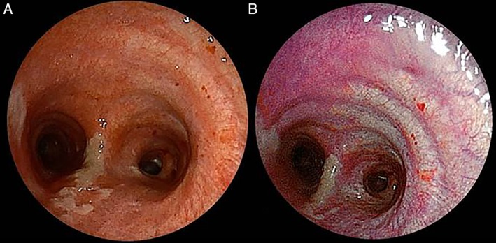
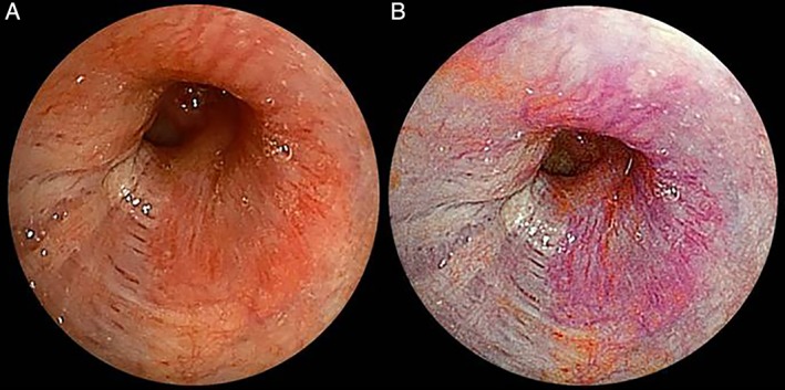
We report two cases of the comparison of diagnosis made with linked color imaging (LCI) and conventional white-light imaging (WLI) on the same patients. In case 1, a 75-year-old man in whom right upper lobectomy with mediastinal lymph node dissection was performed due to lung cancer had signs of bronchitis on postoperative day 8. The LCI demonstrated slight inflammatory changes that were not detectable with the conventional WLI on the tracheal wall. In case 2, in a 61-year-old woman who was diagnosed with adenoid cystic carcinoma, the bronchial wall was checked to confirm the extent of the tumour. The submucosal vascularity and tumour margin on the bronchial mucosa were better visible on LCI than on WLI. We could easily detect the mucosal inflammatory lesion and the malignant lesion with LCI in comparison with conventional WLI. Both mucosal inflammatory and malignant lesions were better visible with LCI in comparison to WLI.

摘要

我们报告了两例针对同一患者使用联动彩色成像(LCI)和传统白光成像(WLI)进行诊断比较的病例。病例1中,一名75岁男性因肺癌接受了右上叶切除及纵隔淋巴结清扫术,术后第8天出现支气管炎迹象。LCI显示气管壁有轻微炎症变化,而传统WLI未检测到这些变化。病例2中,一名61岁女性被诊断为腺样囊性癌,检查支气管壁以确认肿瘤范围。与WLI相比,LCI能更清晰地显示支气管黏膜下的血管分布及肿瘤边缘。与传统WLI相比,使用LCI我们能够轻松检测到黏膜炎症病变和恶性病变。与WLI相比,LCI能更清晰地显示黏膜炎症和恶性病变。

https://cdn.ncbi.nlm.nih.gov/pmc/blobs/03fe/6375225/78c719d4ae3e/RCR2-7-e00399-g002.jpg
https://cdn.ncbi.nlm.nih.gov/pmc/blobs/03fe/6375225/eca1b3fe1b0f/RCR2-7-e00399-g001.jpg
https://cdn.ncbi.nlm.nih.gov/pmc/blobs/03fe/6375225/78c719d4ae3e/RCR2-7-e00399-g002.jpg
https://cdn.ncbi.nlm.nih.gov/pmc/blobs/03fe/6375225/eca1b3fe1b0f/RCR2-7-e00399-g001.jpg
https://cdn.ncbi.nlm.nih.gov/pmc/blobs/03fe/6375225/78c719d4ae3e/RCR2-7-e00399-g002.jpg

相似文献

1
Bronchoscopic observation with linked colour imaging.采用联动彩色成像进行支气管镜观察。
Respirol Case Rep. 2019 Feb 14;7(3):e00399. doi: 10.1002/rcr2.399. eCollection 2019 Apr.
2
Evaluation of Minimal Change Lesions Using Linked Color Imaging in Patients With Nonerosive Reflux Esophagitis.在非糜烂性反流性食管炎患者中使用链接彩色成像评估微小病变
J Clin Gastroenterol. 2022;56(5):405-411. doi: 10.1097/MCG.0000000000001538.
3
Linked color imaging (LCI), a novel image-enhanced endoscopy technology, emphasizes the color of early gastric cancer.联动成像(LCI)是一种新型的图像增强内镜技术,它突出了早期胃癌的颜色。
Endosc Int Open. 2017 Oct;5(10):E1005-E1013. doi: 10.1055/s-0043-117881. Epub 2017 Oct 10.
4
A prospective randomized tandem gastroscopy pilot study of linked color imaging versus white light imaging for detection of upper gastrointestinal lesions.一项关于联合彩色成像与白光成像检测上消化道病变的前瞻性随机串联胃镜试点研究。
J Gastroenterol Hepatol. 2021 Sep;36(9):2562-2567. doi: 10.1111/jgh.15515. Epub 2021 Apr 12.
5
Usefulness of linked color imaging for the detection of obscure early gastric cancer: Multivariate analysis of 508 lesions.链接色成像在检测隐匿性早期胃癌中的作用:508 个病灶的多变量分析。
Dig Endosc. 2022 Jul;34(5):1012-1020. doi: 10.1111/den.14221. Epub 2022 Jan 17.
6
Utility of linked color imaging for endoscopic diagnosis of early gastric cancer.链接色成像在早期胃癌内镜诊断中的应用。
World J Gastroenterol. 2019 Mar 14;25(10):1248-1258. doi: 10.3748/wjg.v25.i10.1248.
7
Efficacy of linked colour imaging in magnifying chromoendoscopy with crystal violet staining: a pilot study.联合色彩成像在放大染色后 chromoendoscopy 中的疗效:一项初步研究。
Int J Colorectal Dis. 2019 Jul;34(7):1341-1344. doi: 10.1007/s00384-019-03323-0. Epub 2019 Jun 5.
8
Diagnostic utility of linked color imaging in the evaluation of colonic mucosal inflammation in ulcerative colitis: a pilot study.链接彩色成像在溃疡性结肠炎结肠黏膜炎症评估中的诊断效用:一项初步研究
Endosc Int Open. 2019 Aug;7(8):E937-E943. doi: 10.1055/a-0810-0398. Epub 2019 Jul 24.
9
Images of laser and light-emitting diode colonoscopy for comparing large colorectal lesion visibility with linked color imaging and white-light imaging.激光和发光二极管结肠镜检查图像比较,用于比较大的结直肠病变的可视性与连接颜色成像和白光成像。
Dig Endosc. 2022 Nov;34(7):1413-1421. doi: 10.1111/den.14370. Epub 2022 Jul 26.
10
Linked Color Imaging Focused on Neoplasm Detection in the Upper Gastrointestinal Tract : A Randomized Trial.链接染色成像在上消化道肿瘤检测中的应用:一项随机试验。
Ann Intern Med. 2021 Jan;174(1):18-24. doi: 10.7326/M19-2561. Epub 2020 Oct 20.

引用本文的文献

1
Primary tracheal schwannoma: Bronchoscopic management of a rare tracheal tumour.原发性气管神经鞘瘤:一种罕见气管肿瘤的支气管镜治疗
Respirol Case Rep. 2024 May 27;12(5):e01390. doi: 10.1002/rcr2.1390. eCollection 2024 May.

本文引用的文献

1
Linked-color imaging improves endoscopic visibility of colorectal nongranular flat lesions.连码显色成像提高结直肠无颗粒平坦病变的内镜可视性。
Gastrointest Endosc. 2017 Oct;86(4):692-697. doi: 10.1016/j.gie.2017.01.044. Epub 2017 Feb 11.
2
Linked color imaging application for improving the endoscopic diagnosis accuracy: a pilot study.链接色成像应用提高内镜诊断准确性的初步研究。
Sci Rep. 2016 Sep 19;6:33473. doi: 10.1038/srep33473.
3
Linked color imaging technology facilitates early detection of flat gastric cancers.联动成像技术有助于早期发现胃扁平癌。
Clin J Gastroenterol. 2015 Dec;8(6):385-9. doi: 10.1007/s12328-015-0612-9. Epub 2015 Nov 11.
4
Postoperative ischemic change in bronchial stumps after primary lung cancer resection.原发性肺癌切除术后支气管残端的缺血性改变
Eur J Cardiothorac Surg. 2006 Jul;30(1):172-6. doi: 10.1016/j.ejcts.2006.03.047. Epub 2006 May 26.
5
High magnification bronchovideoscopy combined with narrow band imaging could detect capillary loops of angiogenic squamous dysplasia in heavy smokers at high risk for lung cancer.高倍支气管镜检查联合窄带成像能够检测出肺癌高危重度吸烟者血管生成性鳞状发育异常的毛细血管袢。
Thorax. 2003 Nov;58(11):989-95. doi: 10.1136/thorax.58.11.989.