Kanzaki Hiromitsu, Takenaka Ryuta, Kawahara Yoshiro, Kawai Daisuke, Obayashi Yuka, Baba Yuki, Sakae Hiroyuki, Gotoda Tatsuhiro, Kono Yoshiyasu, Miura Ko, Iwamuro Masaya, Kawano Seiji, Tanaka Takehiro, Okada Hiroyuki
Department of Gastroenterology and Hepatology, Okayama University Graduate School of Medicine, Dentistry, and Pharmaceutical Sciences, Okayama, Japan.
Department of Gastroenterology, Tsuyama Chuo Hospital, Tsuyama, Japan.
Endosc Int Open. 2017 Oct;5(10):E1005-E1013. doi: 10.1055/s-0043-117881. Epub 2017 Oct 10.
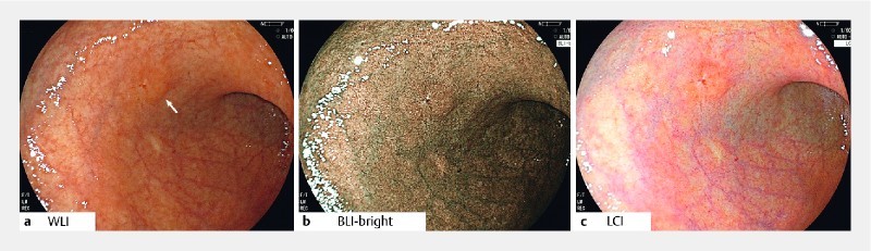
Linked color imaging (LCI) and blue laser imaging (BLI) are novel image-enhanced endoscopy technologies with strong, unique color enhancement. We investigated the efficacy of LCI and BLI-bright compared to conventional white light imaging (WLI) by measuring the color difference between early gastric cancer lesions and the surrounding mucosa.
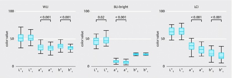
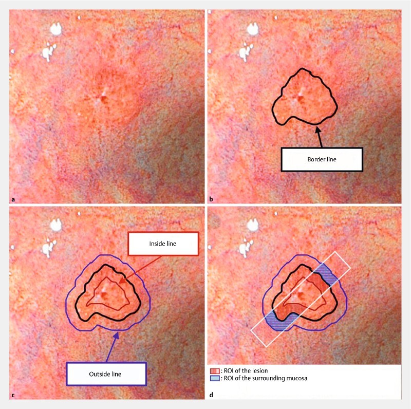
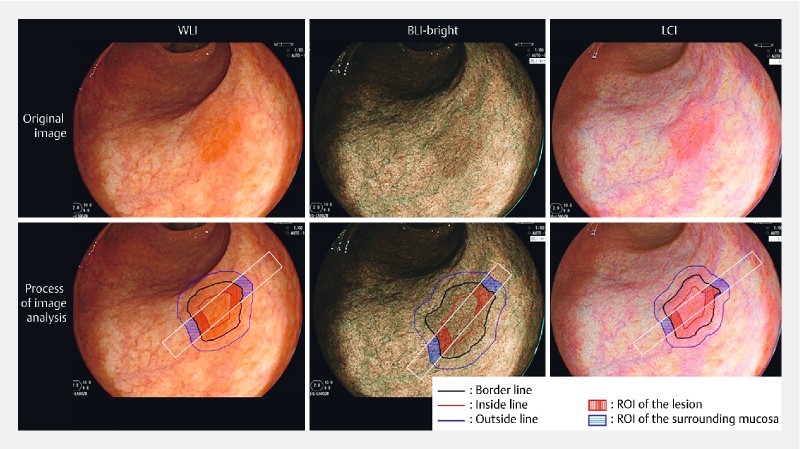
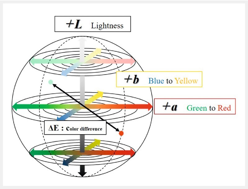
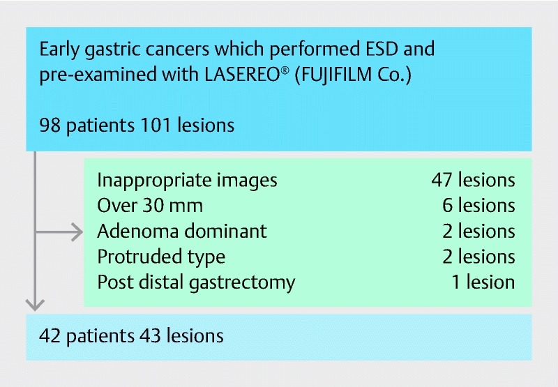
Images of early gastric cancer scheduled for endoscopic submucosal dissection were captured by LCI, BLI-bright, and WLI under the same conditions. Color values of the lesion and surrounding mucosa were defined as the average of the color value in each region of interest. Color differences between the lesion and surrounding mucosa (ΔE) were examined in each mode. The color value was assessed using the CIE Lab* color space (CIE: Commission Internationale d'Eclairage).
We collected images of 43 lesions from 42 patients. Average ΔE values with LCI, BLI-bright, and WLI were 11.02, 5.04, and 5.99, respectively. The ΔE was significantly higher with LCI than with WLI ( < 0.001). Limited to cases of small ΔE with WLI, the ΔE was approximately 3 times higher with LCI than with WLI (7.18 vs. 2.25). The ΔE with LCI was larger when the surrounding mucosa had severe intestinal metaplasia ( = 0.04). The average color value of a lesion and the surrounding mucosa differed. This value did not have a sufficient cut-off point between the lesion and surrounding mucosa to distinguish them, even with LCI.
LCI had a larger ΔE than WLI. It may allow easy recognition and early detection of gastric cancer, even for inexperienced endoscopists.
联动成像(LCI)和蓝光成像(BLI)是新型的图像增强内镜技术,具有强大且独特的色彩增强功能。我们通过测量早期胃癌病变与周围黏膜之间的色差,研究了LCI和BLI-明亮模式相较于传统白光成像(WLI)的有效性。
计划进行内镜黏膜下剥离术的早期胃癌患者在相同条件下分别采用LCI、BLI-明亮模式和WLI进行图像采集。病变及周围黏膜的颜色值定义为每个感兴趣区域颜色值的平均值。在每种模式下检查病变与周围黏膜之间的色差(ΔE)。使用CIE Lab*颜色空间(CIE:国际照明委员会)评估颜色值。
我们收集了42例患者43个病变的图像。LCI、BLI-明亮模式和WLI的平均ΔE值分别为11.02、5.04和5.99。LCI的ΔE显著高于WLI(<0.001)。仅限于WLI色差较小的病例,LCI的ΔE约为WLI的3倍(7.18对2.25)。当周围黏膜存在严重肠化生时,LCI的ΔE更大(=0.04)。病变与周围黏膜的平均颜色值不同。即使使用LCI,该值在病变与周围黏膜之间也没有足够的分界点来区分它们。
LCI的ΔE比WLI大。即使对于经验不足的内镜医师,它也可能有助于轻松识别和早期发现胃癌。