• 文献检索
  • 文档翻译
  • 深度研究
  • 学术资讯
  • Suppr Zotero 插件Zotero 插件
  • 邀请有礼
  • 套餐&价格
  • 历史记录
应用&插件
Suppr Zotero 插件Zotero 插件浏览器插件Mac 客户端Windows 客户端微信小程序
定价
高级版会员购买积分包购买API积分包
服务
文献检索文档翻译深度研究API 文档MCP 服务
关于我们
关于 Suppr公司介绍联系我们用户协议隐私条款
关注我们

Suppr 超能文献

核心技术专利:CN118964589B侵权必究
粤ICP备2023148730 号-1Suppr @ 2026

文献检索

告别复杂PubMed语法,用中文像聊天一样搜索,搜遍4000万医学文献。AI智能推荐,让科研检索更轻松。

立即免费搜索

文件翻译

保留排版,准确专业,支持PDF/Word/PPT等文件格式,支持 12+语言互译。

免费翻译文档

深度研究

AI帮你快速写综述,25分钟生成高质量综述,智能提取关键信息,辅助科研写作。

立即免费体验

影响光学相干断层扫描视盘周围脉络膜厚度的因素:一项多中心研究。

Factors Influencing Optical Coherence Tomography Peripapillary Choroidal Thickness: A Multicenter Study.

机构信息

Devers Eye Institute, Optic Nerve Head Research Laboratory, Legacy Research Institute, Portland, Oregon, United States.

Department of Ophthalmology, Second Xiangya Hospital, Central South University, Changsha, Hunan Province, People's Republic of China.

出版信息

Invest Ophthalmol Vis Sci. 2019 Feb 1;60(2):795-806. doi: 10.1167/iovs.18-25407.

DOI:10.1167/iovs.18-25407
PMID:30811523
原文链接:https://pmc.ncbi.nlm.nih.gov/articles/PMC6392476/
Abstract

PURPOSE

To quantify peripapillary choroidal thickness (PCT) and the factors that influence it in healthy participants who represent the racial and ethnic composition of the U.S. population.

METHODS

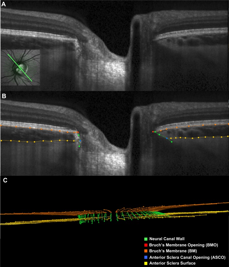
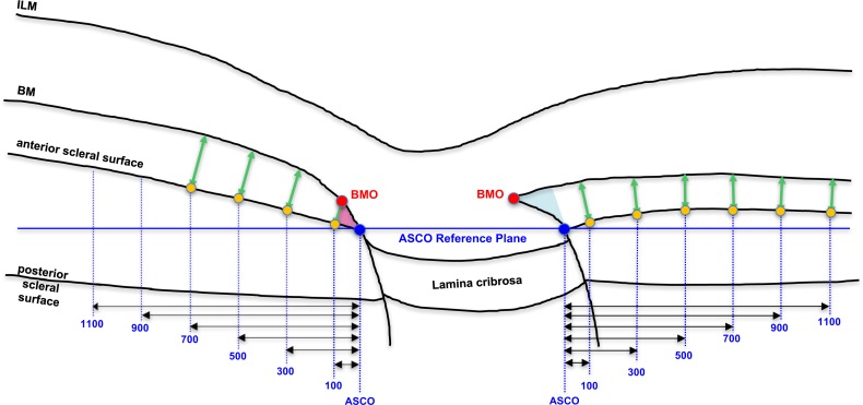
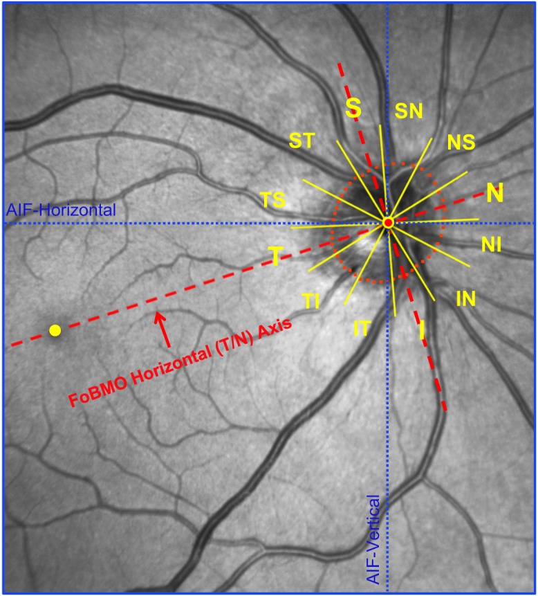
A total of 362 healthy participants underwent optical coherence tomography (OCT) enhanced depth imaging of the optic nerve head with a 24 radial B-scan pattern aligned to the fovea to Bruch's membrane opening axis. Bruch's membrane, anterior scleral canal opening (ASCO), and the anterior scleral surface were manually segmented. PCT was measured at 100, 300, 500, 700, 900, and 1100 μm from the ASCO globally and within 12 clock-hour sectors. The effects of age, axial length, intraocular pressure, ethnicity, sex, sector, and ASCO area on PCT were assessed by ANOVA and univariable and multivariable regressions.

RESULTS

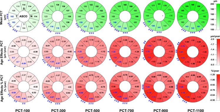
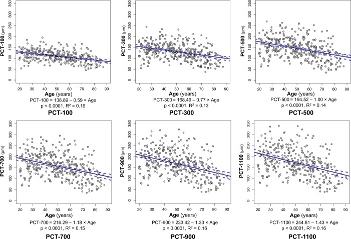
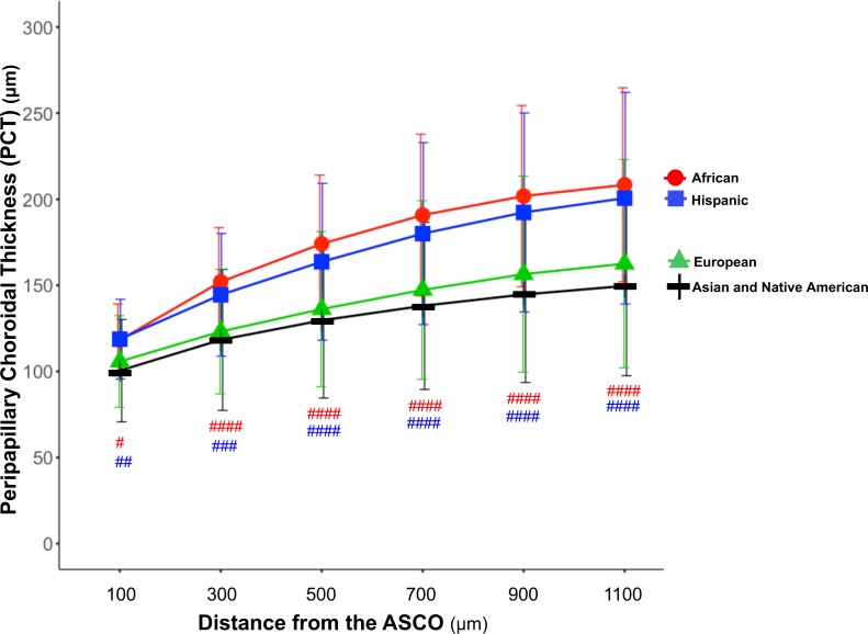
Globally, PCT was thicker further from the ASCO border and thinner with older age, longer axial length, larger ASCO area, European descent, and female sex. Among these effectors, age and axial length explained the greatest proportion of variance. The rate of age-related decline increased further from the ASCO border. Sectorally, the inferior-temporal sectors were thinnest (10.7%-20.0% thinner than the thickest sector) and demonstrated a higher rate of age-related loss (from 15.6% to 20.7% faster) at each ASCO distance.

CONCLUSIONS

In healthy eyes, PCT was thinnest in the inferior temporal sectors and thinner PCT was associated with older age, European descent, longer axial length, larger ASCO area, and female sex. Among these associations, age had the strongest influence, and its effect was greatest within the inferior temporal sectors.

摘要

目的

定量测量代表美国人口种族和民族构成的健康参与者的视盘周围脉络膜厚度(PCT)及其影响因素。

方法

共有 362 名健康参与者接受了视神经头光学相干断层扫描(OCT)增强深度成像检查,采用 24 条与黄斑至脉络膜开口轴对准的放射状 B 扫描模式。手动分割脉络膜、前巩膜管开口(ASCO)和前巩膜表面。在距 ASCO 全局和 12 个时钟小时扇区 100、300、500、700、900 和 1100μm 处测量 PCT。通过 ANOVA 和单变量及多变量回归评估年龄、眼轴长度、眼内压、种族、性别、扇区和 ASCO 面积对 PCT 的影响。

结果

全局而言,PCT 距 ASCO 边界越远越厚,年龄越大、眼轴越长、ASCO 面积越大、欧洲血统和女性性别越薄。在这些影响因素中,年龄和眼轴长度解释了最大的方差比例。从 ASCO 边界向外,与年龄相关的下降速度进一步增加。在扇形方面,下颞部最薄(比最厚的扇形薄 10.7%-20.0%),并且在每个 ASCO 距离处,与年龄相关的损失速度更快(从 15.6%增加到 20.7%)。

结论

在健康的眼睛中,下颞部的 PCT 最薄,较薄的 PCT 与年龄较大、欧洲血统、眼轴较长、ASCO 面积较大和女性性别有关。在这些关联中,年龄的影响最大,其影响在颞下区最大。

https://cdn.ncbi.nlm.nih.gov/pmc/blobs/bcf4/6392476/e9f16d8f58eb/i1552-5783-60-2-795-f07.jpg
https://cdn.ncbi.nlm.nih.gov/pmc/blobs/bcf4/6392476/50ff704bbfce/i1552-5783-60-2-795-f01.jpg
https://cdn.ncbi.nlm.nih.gov/pmc/blobs/bcf4/6392476/93efe18cc65d/i1552-5783-60-2-795-f02.jpg
https://cdn.ncbi.nlm.nih.gov/pmc/blobs/bcf4/6392476/186da0168560/i1552-5783-60-2-795-f03.jpg
https://cdn.ncbi.nlm.nih.gov/pmc/blobs/bcf4/6392476/56ec69ab6129/i1552-5783-60-2-795-f04.jpg
https://cdn.ncbi.nlm.nih.gov/pmc/blobs/bcf4/6392476/62ddc42afdae/i1552-5783-60-2-795-f05.jpg
https://cdn.ncbi.nlm.nih.gov/pmc/blobs/bcf4/6392476/7ed167be3dcd/i1552-5783-60-2-795-f06.jpg
https://cdn.ncbi.nlm.nih.gov/pmc/blobs/bcf4/6392476/e9f16d8f58eb/i1552-5783-60-2-795-f07.jpg

相似文献

1
Factors Influencing Optical Coherence Tomography Peripapillary Choroidal Thickness: A Multicenter Study.影响光学相干断层扫描视盘周围脉络膜厚度的因素:一项多中心研究。
Invest Ophthalmol Vis Sci. 2019 Feb 1;60(2):795-806. doi: 10.1167/iovs.18-25407.
2
Factors Influencing Central Lamina Cribrosa Depth: A Multicenter Study.影响中枢层视盘深度的因素:一项多中心研究。
Invest Ophthalmol Vis Sci. 2018 May 1;59(6):2357-2370. doi: 10.1167/iovs.17-23456.
3
Assessment of Open-Angle Glaucoma Peripapillary and Macular Choroidal Thickness Using Swept-Source Optical Coherence Tomography (SS-OCT).使用扫频光学相干断层扫描(SS-OCT)评估开角型青光眼视乳头周围和黄斑脉络膜厚度。
PLoS One. 2016 Jun 16;11(6):e0157333. doi: 10.1371/journal.pone.0157333. eCollection 2016.
4
Peripapillary Scleral Bowing Increases with Age and Is Inversely Associated with Peripapillary Choroidal Thickness in Healthy Eyes.周边巩膜弓随年龄增长而增加,与健康眼中的脉络膜厚度呈负相关。
Am J Ophthalmol. 2020 Sep;217:91-103. doi: 10.1016/j.ajo.2020.03.050. Epub 2020 Apr 13.
5
Peripapillary choroidal thickness variation with age and race in normal eyes.正常眼视乳头周围脉络膜厚度随年龄和种族的变化
Invest Ophthalmol Vis Sci. 2015 Feb 24;56(3):1872-9. doi: 10.1167/iovs.14-16179.
6
Variation of the axial location of Bruch's membrane opening with age, choroidal thickness, and race.Bruch 膜开口的轴向位置随年龄、脉络膜厚度和种族的变化。
Invest Ophthalmol Vis Sci. 2014 Mar 28;55(3):2004-9. doi: 10.1167/iovs.13-12937.
7
Evaluation of peripapillary choroidal distribution in children by enhanced depth imaging optical coherence tomography.通过增强深度成像光学相干断层扫描评估儿童视乳头周围脉络膜分布情况。
BMC Ophthalmol. 2018 Jul 13;18(1):173. doi: 10.1186/s12886-018-0839-6.
8
Peripapillary Pachychoroid in Nonarteritic Anterior Ischemic Optic Neuropathy.非动脉炎性前部缺血性视神经病变中的视乳头周围厚脉络膜
Invest Ophthalmol Vis Sci. 2016 Sep 1;57(11):4679-85. doi: 10.1167/iovs.16-19315.
9
OCT Optic Nerve Head Morphology in Myopia IV: Neural Canal Scleral Flange Remodeling in Highly Myopic Eyes.OCT 视神经头形态学在近视 IV 中的研究:高度近视眼中的神经管巩膜脊重塑。
Am J Ophthalmol. 2024 May;261:141-164. doi: 10.1016/j.ajo.2024.01.023. Epub 2024 Feb 3.
10
Peripapillary choroidal vascular layers: the Beijing Eye Study.视乳头周围脉络膜血管层:北京眼病研究
Acta Ophthalmol. 2017 Sep;95(6):619-628. doi: 10.1111/aos.13379. Epub 2017 Mar 2.

引用本文的文献

1
Diagnostic Performance for Detection of Glaucomatous Structural Damage Using Pixelwise Analysis of Retinal Thickness Measurements.基于视网膜厚度测量的像素分析对青光眼结构损伤的检测诊断性能。
Invest Ophthalmol Vis Sci. 2024 Oct 1;65(12):17. doi: 10.1167/iovs.65.12.17.
2
Long-Term Changes in Lamina Cribrosa Curvature Index After Trabeculectomy in Glaucomatous Eyes.青光眼患者小梁切除术后板层巩膜嵴曲率指数的长期变化。
Invest Ophthalmol Vis Sci. 2024 Sep 3;65(11):3. doi: 10.1167/iovs.65.11.3.
3
Choroidal and Retinal Thicknesses in Healthy Eyes Measured with Ultra-Wide-Field Optical Coherence Tomography.

本文引用的文献

1
Detection of Bruch's Membrane Opening in Healthy Individuals and Glaucoma Patients with and without High Myopia.健康个体以及伴有和不伴有高度近视的青光眼患者中 Bruch 膜开口的检测。
Ophthalmology. 2018 Oct;125(10):1537-1546. doi: 10.1016/j.ophtha.2018.04.031. Epub 2018 Jun 19.
2
Factors Influencing Central Lamina Cribrosa Depth: A Multicenter Study.影响中枢层视盘深度的因素:一项多中心研究。
Invest Ophthalmol Vis Sci. 2018 May 1;59(6):2357-2370. doi: 10.1167/iovs.17-23456.
3
Diagnostic capability of optic nerve head rim width and retinal nerve fiber thickness in open-angle glaucoma.
使用超广角光学相干断层扫描测量健康眼睛的脉络膜和视网膜厚度。
Diagnostics (Basel). 2024 May 28;14(11):1114. doi: 10.3390/diagnostics14111114.
4
Heterogenous thinning of peripapillary tissues occurs early during high myopia development in juvenile tree shrews.青少年树鼩高度近视发展早期出现视盘周围组织异质性变薄。
Exp Eye Res. 2024 Mar;240:109824. doi: 10.1016/j.exer.2024.109824. Epub 2024 Feb 7.
5
OCT Optic Nerve Head Morphology in Myopia IV: Neural Canal Scleral Flange Remodeling in Highly Myopic Eyes.OCT 视神经头形态学在近视 IV 中的研究:高度近视眼中的神经管巩膜脊重塑。
Am J Ophthalmol. 2024 May;261:141-164. doi: 10.1016/j.ajo.2024.01.023. Epub 2024 Feb 3.
6
Deep Optic Nerve Head Morphology in Tilted Disc Syndrome and Its Clinical Implication on Visual Damage.倾斜盘综合征的视神经头形态及其对视功能损害的临床意义
Invest Ophthalmol Vis Sci. 2023 Oct 3;64(13):10. doi: 10.1167/iovs.64.13.10.
7
Optical Coherence Tomographic Optic Nerve Head Morphology in Myopia III: The Exposed Neural Canal Region in Healthy Eyes-Implications for High Myopia.近视 III 眼中的光学相干断层扫描视神经头形态:健康眼中暴露的神经管区域 - 对高度近视的影响。
Am J Ophthalmol. 2024 Feb;258:55-75. doi: 10.1016/j.ajo.2023.08.012. Epub 2023 Sep 5.
8
Differences in Central and Peripheral Choroidal Thickness among the Subtypes of Age-Related Macular Degeneration in an Asian Population.亚洲人群年龄相关性黄斑变性各亚型中央和周边脉络膜厚度的差异
J Clin Med. 2023 Aug 18;12(16):5364. doi: 10.3390/jcm12165364.
9
OCT Optic Nerve Head Morphology in Myopia II: Peri-Neural Canal Scleral Bowing and Choroidal Thickness in High Myopia-An American Ophthalmological Society Thesis.OCT 视神经头形态在近视 II 中的研究:高近视中的神经周围管巩膜弓和脉络膜厚度——美国眼科学会论文。
Am J Ophthalmol. 2023 Aug;252:225-252. doi: 10.1016/j.ajo.2023.03.002. Epub 2023 Mar 10.
10
Deep Optic Nerve Head Structures Associated With Increasing Axial Length in Healthy Myopic Eyes of Moderate Axial Length.健康中度近视眼轴增长相关的深层视神经头结构。
Am J Ophthalmol. 2023 May;249:156-166. doi: 10.1016/j.ajo.2023.01.003. Epub 2023 Jan 14.
视神经乳头边缘宽度和视网膜神经纤维厚度在开角型青光眼中的诊断能力
Eur J Ophthalmol. 2018 Jul;28(4):459-464. doi: 10.1177/1120672117750057. Epub 2018 Mar 19.
4
Detecting Glaucomatous Progression With a Region-of-Interest Approach on Optical Coherence Tomography: A Signal-to-Noise Evaluation.利用光学相干断层扫描的感兴趣区域方法检测青光眼进展:信噪比评估
Transl Vis Sci Technol. 2018 Feb 28;7(1):19. doi: 10.1167/tvst.7.1.19. eCollection 2018 Feb.
5
Choroidal Microvasculature Dropout is Not Associated With Myopia, But is Associated With Glaucoma.脉络膜微血管缺失与近视无关,但与青光眼有关。
J Glaucoma. 2018 Feb;27(2):189-196. doi: 10.1097/IJG.0000000000000859.
6
Detecting Structural Progression in Glaucoma with Optical Coherence Tomography.利用光学相干断层扫描技术检测青光眼的结构进展
Ophthalmology. 2017 Dec;124(12S):S57-S65. doi: 10.1016/j.ophtha.2017.07.015.
7
Determinants and Characteristics of Bruch's Membrane Opening and Bruch's Membrane Opening-Minimum Rim Width in a Normal Japanese Population.日本正常人群中布鲁赫膜开口及布鲁赫膜开口最小边缘宽度的决定因素和特征
Invest Ophthalmol Vis Sci. 2017 Aug 1;58(10):4106-4113. doi: 10.1167/iovs.17-22057.
8
Parapapillary Choroidal Microvasculature Dropout in Glaucoma: A Comparison between Optical Coherence Tomography Angiography and Indocyanine Green Angiography.青光眼的神经旁脉络膜毛细血管缺失:光学相干断层扫描血管造影与吲哚菁绿血管造影的比较。
Ophthalmology. 2017 Aug;124(8):1209-1217. doi: 10.1016/j.ophtha.2017.03.039. Epub 2017 Apr 19.
9
Measurements of the parapapillary atrophy zones in en face optical coherence tomography images.在正面光学相干断层扫描图像中对视乳头周围萎缩区的测量。
PLoS One. 2017 Apr 17;12(4):e0175347. doi: 10.1371/journal.pone.0175347. eCollection 2017.
10
Deep Retinal Layer Microvasculature Dropout Detected by the Optical Coherence Tomography Angiography in Glaucoma.光学相干断层扫描血管造影术检测青光眼患者视网膜深层微血管缺失
Ophthalmology. 2016 Dec;123(12):2509-2518. doi: 10.1016/j.ophtha.2016.09.002. Epub 2016 Oct 18.