Department Immunology, Institut Recerca Hospital de La Santa Creu i Sant Pau, Barcelona, Spain.
Pleural Medicine Unit, Department Internal Medicine, Hospital Universitari Arnau de Vilanova, Lleida, Spain.
Sci Rep. 2019 Feb 28;9(1):2996. doi: 10.1038/s41598-018-35840-3.
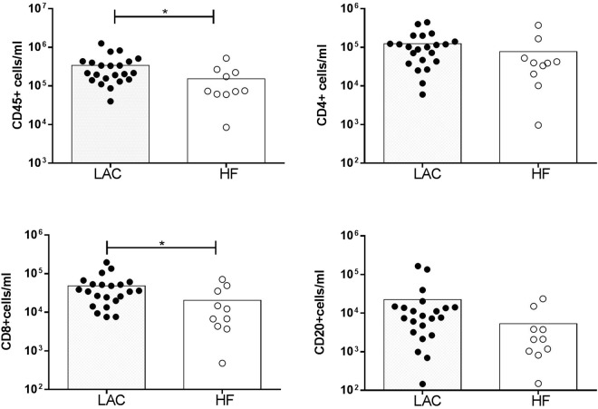
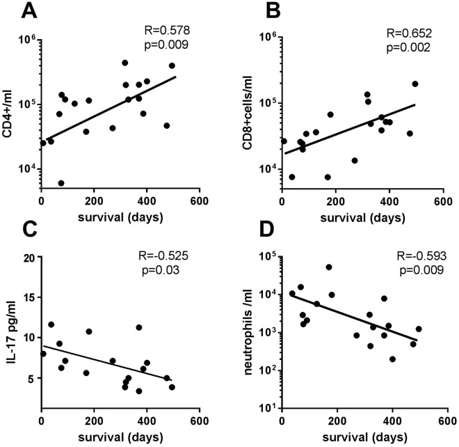
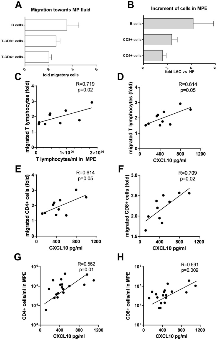
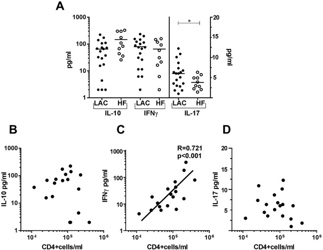
The presence of leukocyte subpopulations in malignant pleural effusions (MPEs) can have a different impact on tumor cell proliferation and vascular leakiness, their analysis can help to understand the metastatic microenvironment. We analyzed the relationship between the leukocyte subpopulation counts per ml of pleural fluid and the tumor cell count, molecular phenotype of lung adenocarcinoma (LAC), time from cancer diagnosis and previous oncologic therapy. We also evaluated the leukocyte composition of MPEs as a biomarker of prognosis. We determined CD4+ T, CD8+ T and CD20+ B cells, monocytes and neutrophils per ml in pleural effusions of 22 LAC and 10 heart failure (HF) patients by flow cytometry. Tumor cells were identified by morphology and CD326 expression. IFNγ, IL-10 and IL-17, and chemokines were determined by ELISAs and migratory response to pleural fluids by transwell assays. MPEs from LAC patients had more CD8+ T lymphocytes and a tendency to more CD4+ T and CD20+ B lymphocytes than HF-related fluids. However, no correlation was found between lymphocytes and tumor cells. In those MPEs which were detected >1 month from LAC diagnosis, there was a negative correlation between pleural tumor cells and CD8+ T lymphocytes. CXCL10 was responsible for the attraction of CD20+ B, CD4+ T and CD8+ T lymphocytes in malignant fluids. Concentrations of IL-17 were higher in MPEs than in HF-related effusions. Survival after MPE diagnosis correlated positively with CD4+ T and CD8+ T lymphocytes, but negatively with neutrophils and IL-17 levels. In conclusion, lymphocyte enrichment in MPEs from LAC patients is mostly due to local migration and increases patient survival.
恶性胸腔积液(MPE)中白细胞亚群的存在可能对肿瘤细胞增殖和血管通透性产生不同的影响,其分析有助于了解转移微环境。我们分析了胸腔液中白细胞亚群计数与肿瘤细胞计数、肺腺癌(LAC)分子表型、从癌症诊断到先前肿瘤治疗的时间以及先前肿瘤治疗之间的关系。我们还评估了 MPE 中的白细胞组成作为预后的生物标志物。我们通过流式细胞术确定了 22 例 LAC 和 10 例心力衰竭(HF)患者胸腔积液中的每毫升 CD4+T、CD8+T 和 CD20+B 细胞、单核细胞和中性粒细胞。通过形态学和 CD326 表达鉴定肿瘤细胞。通过 ELISA 测定 IFNγ、IL-10 和 IL-17 以及趋化因子,并通过 Transwell 测定胸腔液的迁移反应。与 HF 相关的液体相比,LAC 患者的 MPE 具有更多的 CD8+T 淋巴细胞,并且倾向于具有更多的 CD4+T 和 CD20+B 淋巴细胞。然而,淋巴细胞与肿瘤细胞之间没有相关性。在从 LAC 诊断超过 1 个月检测到的那些 MPE 中,胸腔肿瘤细胞与 CD8+T 淋巴细胞之间存在负相关。CXCL10 负责吸引恶性液中的 CD20+B、CD4+T 和 CD8+T 淋巴细胞。MPE 中的 IL-17 浓度高于 HF 相关的胸腔积液。MPE 诊断后的生存与 CD4+T 和 CD8+T 淋巴细胞呈正相关,但与中性粒细胞和 IL-17 水平呈负相关。总之,LAC 患者 MPE 中淋巴细胞的富集主要归因于局部迁移并增加了患者的生存。