Department of Mechanical Engineering, Johns Hopkins University, Baltimore, Maryland.
Department of Biomedical Engineering, University of Arkansas, Fayetteville, Arkansas.
Cancer Res. 2019 Apr 15;79(8):2054-2064. doi: 10.1158/0008-5472.CAN-18-2732. Epub 2019 Feb 28.
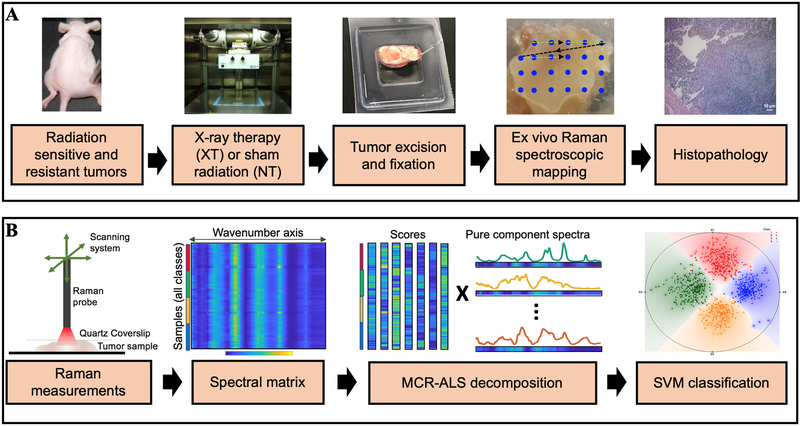
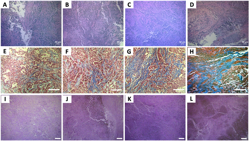
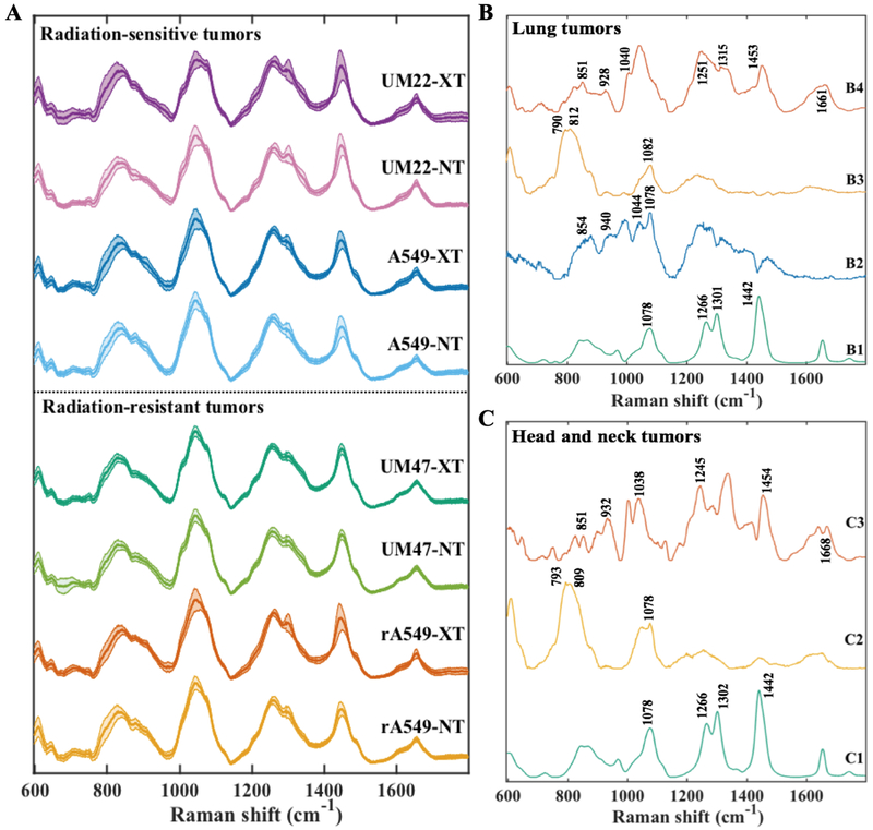
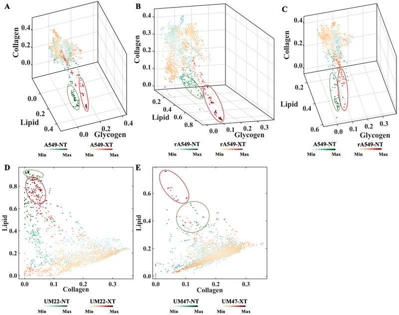
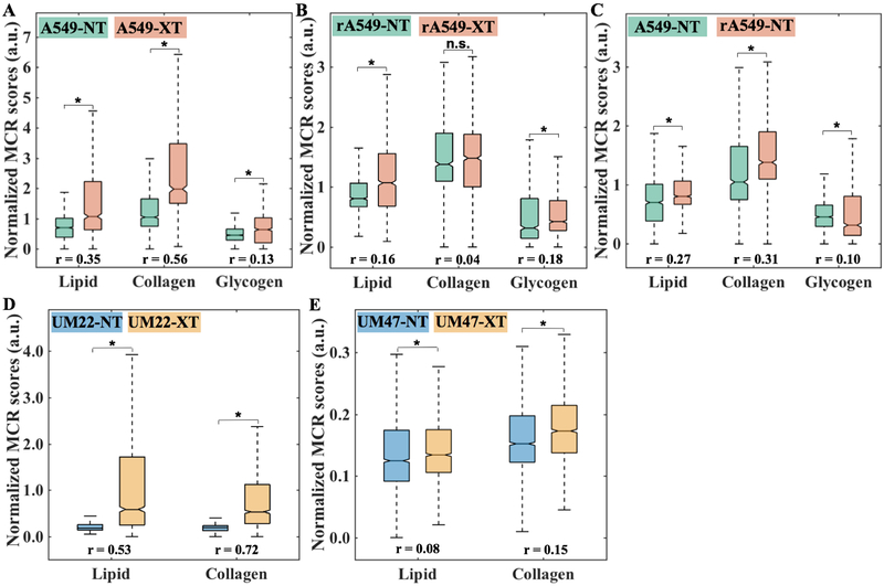
Delay in the assessment of tumor response to radiotherapy continues to pose a major challenge to quality of life for patients with nonresponsive tumors. Here, we exploited label-free Raman spectroscopic mapping to elucidate radiation-induced biomolecular changes in tumors and uncovered latent microenvironmental differences between treatment-resistant and -sensitive tumors. We used isogenic radiation-resistant and -sensitive A549 human lung cancer cells and human head and neck squamous cell carcinoma (HNSCC) cell lines (UM-SCC-47 and UM-SCC-22B, respectively) to grow tumor xenografts in athymic nude mice and demonstrated the molecular specificity and quantitative nature of Raman spectroscopic tissue assessments. Raman spectra obtained from untreated and treated tumors were subjected to chemometric analysis using multivariate curve resolution-alternating least squares (MCR-ALS) and support vector machine (SVM) to quantify biomolecular differences in the tumor microenvironment. The Raman measurements revealed significant and reliable differences in lipid and collagen content postradiation in the tumor microenvironment, with consistently greater changes observed in the radiation-sensitive tumors. In addition to accurately evaluating tumor response to therapy, the combination of Raman spectral markers potentially offers a route to predicting response in untreated tumors prior to commencing treatment. Combined with its noninvasive nature, our findings provide a rationale for studies using Raman spectroscopy, with the ultimate goal of clinical translation for patient stratification and guiding adaptation of radiotherapy during the course of treatment. SIGNIFICANCE: These findings highlight the sensitivity of label-free Raman spectroscopy to changes induced by radiotherapy and indicate the potential to predict radiation resistance prior to commencing therapy.
肿瘤对放射治疗的反应评估延迟仍然是对无反应性肿瘤患者生活质量的主要挑战。在这里,我们利用无标记拉曼光谱图谱来阐明肿瘤中放射诱导的生物分子变化,并揭示了治疗抵抗性和敏感性肿瘤之间潜在的微观环境差异。我们使用同源性的放射抵抗性和敏感性 A549 人肺癌细胞和人头颈鳞状细胞癌(HNSCC)细胞系(分别为 UM-SCC-47 和 UM-SCC-22B)在裸鼠中生长肿瘤异种移植物,并证明了拉曼光谱组织评估的分子特异性和定量性质。对未处理和处理的肿瘤进行拉曼光谱采集,并使用多变量曲线分辨交替最小二乘法(MCR-ALS)和支持向量机(SVM)对其进行化学计量分析,以量化肿瘤微环境中的生物分子差异。拉曼测量结果显示,在肿瘤微环境中,辐射后脂质和胶原含量存在显著且可靠的差异,在辐射敏感性肿瘤中观察到的变化更大。除了准确评估肿瘤对治疗的反应外,拉曼光谱标志物的组合还可能提供一种在开始治疗之前预测未处理肿瘤反应的方法。结合其非侵入性,我们的研究结果为使用拉曼光谱进行研究提供了依据,最终目标是为患者分层和指导治疗过程中的放射治疗适应提供临床转化。意义:这些发现强调了无标记拉曼光谱对放射治疗诱导变化的敏感性,并表明在开始治疗之前预测放射抵抗的潜力。