Division of Anatomical Science, Department of Functional Morphology, Nihon University School of Medicine, Tokyo
Cellular and Molecular Toxicology Division, National Center for Biological Safety and Research, National Institute of Health Science, Kawasaki, Japan.
Haematologica. 2019 Oct;104(10):1995-2005. doi: 10.3324/haematol.2018.209551. Epub 2019 Feb 28.
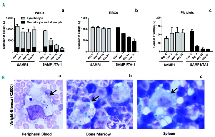
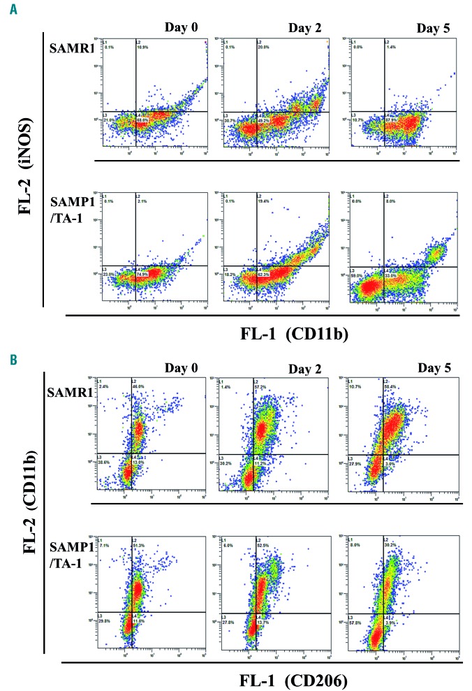
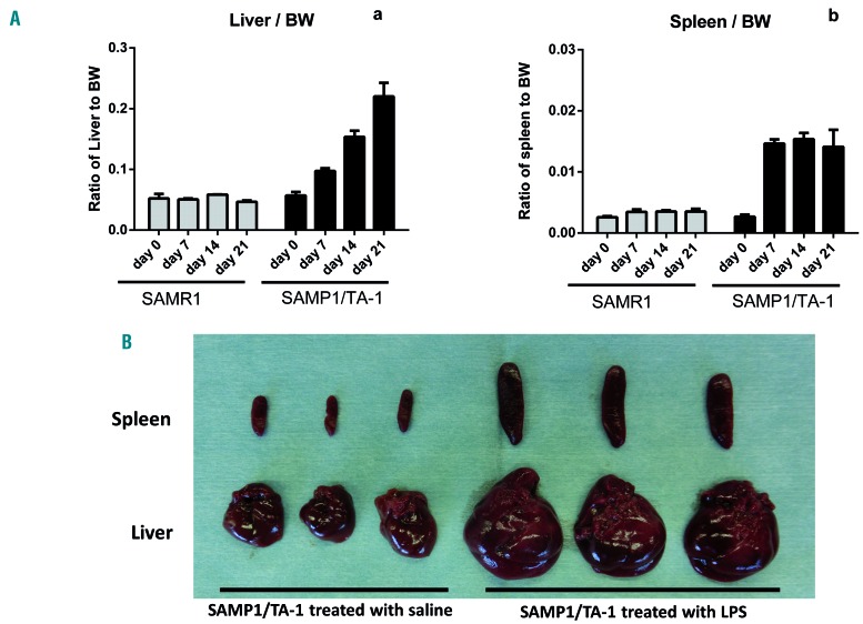
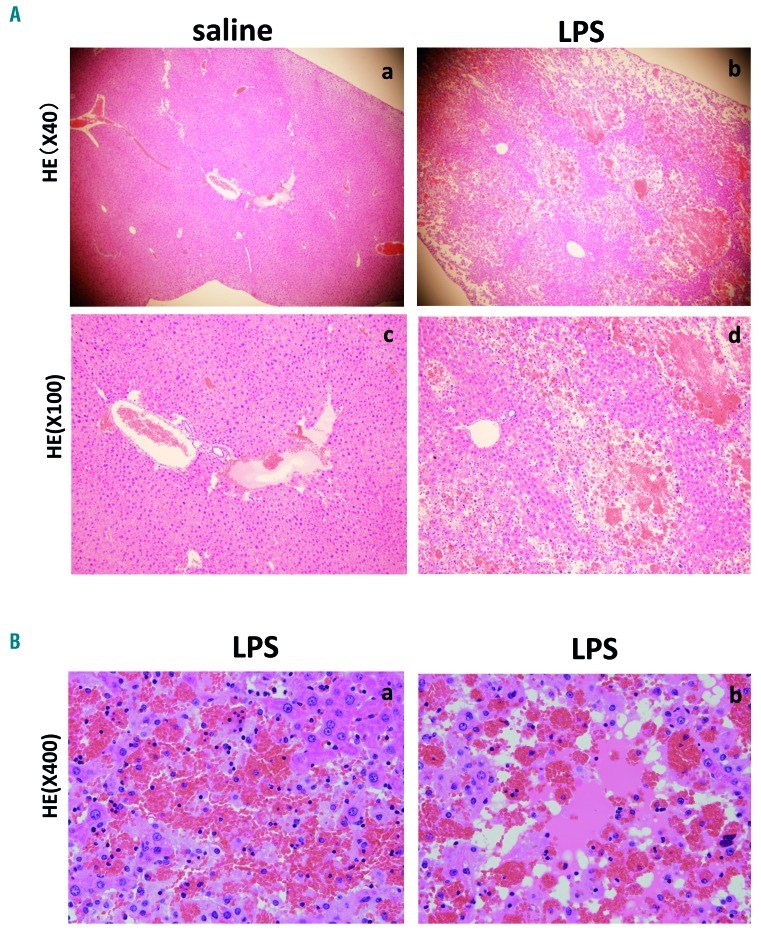
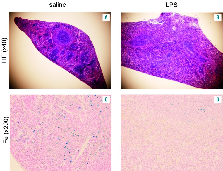
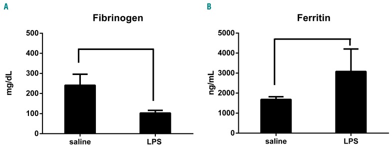
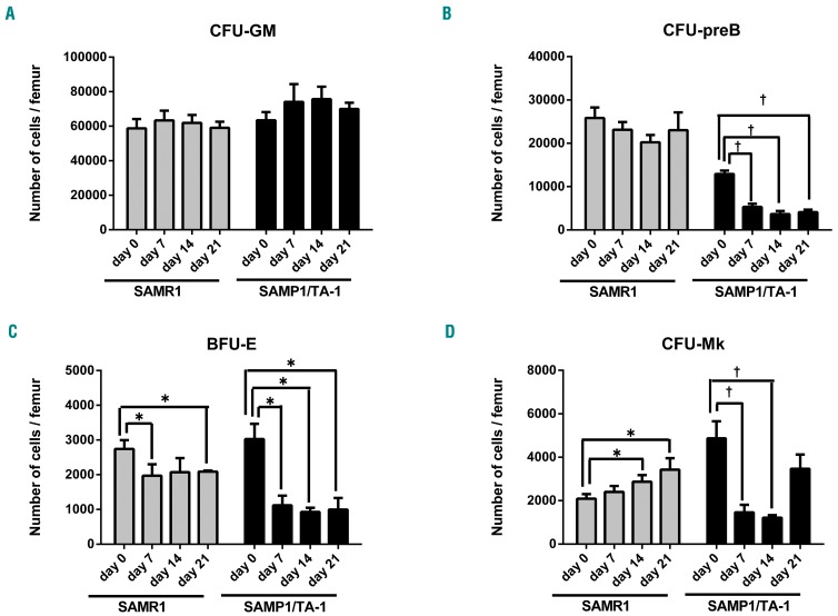
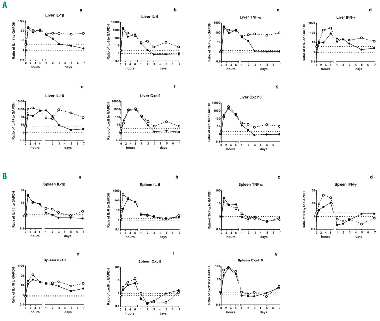
Hemophagocytic lymphohistiocytosis is a life-threatening systemic hyperinflammatory disorder with primary and secondary forms. Primary hemophagocytic lymphohistiocytosis is associated with inherited defects in various genes that affect the immunological cytolytic pathway. Secondary hemophagocytic lymphohistiocytosis is not inherited, but complicates various medical conditions including infections, autoinflammatory/autoimmune diseases, and malignancies. When senescence-accelerated mice (SAMP1/TA-1) with latent deterioration of immunological function and senescence-resistant control mice (SAMR1) were treated repeatedly with lipopolysaccharide, SAMP1/TA-1 mice displayed the clinicopathological features of hemophagocytic lymphohistiocytosis such as hepatosplenomegaly, pancytopenia, hypofibrinogenemia, hyperferritinemia, and hemophagocytosis. SAMR1 mice showed no features of hemophagocytic lymphohistiocytosis. Lipopolysaccharide induced upregulation of proinflammatory cytokines such as interleukin-1β, interleukin-6, tumor necrosis factor-α, and interferon-γ, and interferon-γ-inducible chemokines such as c-x-c motif chemokine ligands 9 and 10 in the liver and spleen in both SAMP1/TA-1 and SAMR1 mice. However, upregulation of proinflammatory cytokines and interferon-γ-inducible chemokines in the liver persisted for longer in SAMP1/TA-1 mice than in SAMR1 mice. In addition, the magnitude of upregulation of interferon-γ in the liver and spleen after lipopolysaccharide treatment was greater in SAMP1/TA-1 mice than in SAMR1 mice. Furthermore, lipopolysaccharide treatment led to a prolonged increase in the proportion of peritoneal M1 macrophages and simultaneously to a decrease in the proportion of M2 macrophages in SAMP1/TA-1 mice compared with SAMR1 mice. Lipopolysaccharide appeared to induce a hyperinflammatory reaction and prolonged inflammation in SAMP1/TA-1 mice, resulting in features of secondary hemophagocytic lymphohistiocytosis. Thus, SAMP1/TA-1 mice represent a useful mouse model to investigate the pathogenesis of bacterial infection-associated secondary hemophagocytic lymphohistiocytosis.
噬血细胞性淋巴组织细胞增生症是一种危及生命的全身性炎症过度活跃性疾病,分为原发性和继发性两种。原发性噬血细胞性淋巴组织细胞增生症与影响免疫细胞溶解途径的各种基因的遗传缺陷有关。继发性噬血细胞性淋巴组织细胞增生症不是遗传性的,但会并发各种疾病,包括感染、自身炎症/自身免疫性疾病和恶性肿瘤。当免疫功能潜伏恶化的快速老化小鼠(SAMP1/TA-1)和免疫衰老抵抗对照小鼠(SAMR1)反复用脂多糖处理时,SAMP1/TA-1 小鼠表现出噬血细胞性淋巴组织细胞增生症的临床病理特征,如肝脾肿大、全血细胞减少、低纤维蛋白原血症、高铁蛋白血症和噬血现象。SAMR1 小鼠没有噬血细胞性淋巴组织细胞增生症的特征。脂多糖诱导 SAMP1/TA-1 和 SAMR1 小鼠肝脏和脾脏中促炎细胞因子(如白细胞介素-1β、白细胞介素-6、肿瘤坏死因子-α和干扰素-γ)和干扰素-γ诱导的趋化因子(如 C-X-C 基序趋化因子配体 9 和 10)的上调。然而,SAMP1/TA-1 小鼠肝脏中促炎细胞因子和干扰素-γ诱导的趋化因子的上调持续时间长于 SAMR1 小鼠。此外,脂多糖处理后 SAMP1/TA-1 小鼠肝脏和脾脏中干扰素-γ的上调幅度大于 SAMR1 小鼠。此外,脂多糖处理导致 SAMP1/TA-1 小鼠腹腔 M1 巨噬细胞比例持续增加,同时 M2 巨噬细胞比例下降。与 SAMR1 小鼠相比,脂多糖似乎在 SAMP1/TA-1 小鼠中诱导了过度的炎症反应和炎症的持续存在,导致了继发性噬血细胞性淋巴组织细胞增生症的特征。因此,SAMP1/TA-1 小鼠代表了一种有用的小鼠模型,可以研究细菌感染相关的继发性噬血细胞性淋巴组织细胞增生症的发病机制。