• 文献检索
  • 文档翻译
  • 深度研究
  • 学术资讯
  • Suppr Zotero 插件Zotero 插件
  • 邀请有礼
  • 套餐&价格
  • 历史记录
应用&插件
Suppr Zotero 插件Zotero 插件浏览器插件Mac 客户端Windows 客户端微信小程序
定价
高级版会员购买积分包购买API积分包
服务
文献检索文档翻译深度研究API 文档MCP 服务
关于我们
关于 Suppr公司介绍联系我们用户协议隐私条款
关注我们

Suppr 超能文献

核心技术专利:CN118964589B侵权必究
粤ICP备2023148730 号-1Suppr @ 2026

文献检索

告别复杂PubMed语法,用中文像聊天一样搜索,搜遍4000万医学文献。AI智能推荐,让科研检索更轻松。

立即免费搜索

文件翻译

保留排版,准确专业,支持PDF/Word/PPT等文件格式,支持 12+语言互译。

免费翻译文档

深度研究

AI帮你快速写综述,25分钟生成高质量综述,智能提取关键信息,辅助科研写作。

立即免费体验

小鼠中该基因单拷贝缺失的眼部表型后果。 (括号内内容原文缺失,无法完整翻译)

Ocular phenotypic consequences of a single copy deletion of the gene ( ) in mice.

作者信息

Kim Soohyun, Thomasy Sara M, Raghunathan Vijay Krishna, Teixeira Leandro B C, Moshiri Ala, FitzGerald Paul, Murphy Christopher J

机构信息

Department of Surgical and Radiological Sciences, School of Veterinary Medicine, University of California Davis, Davis, CA.

Department of Ophthalmology & Vision Science, School of Medicine, School of Medicine, University of California Davis, Davis, CA.

出版信息

Mol Vis. 2019 Feb 17;25:129-142. eCollection 2019.

PMID:30820148
原文链接:https://pmc.ncbi.nlm.nih.gov/articles/PMC6382475/
Abstract

PURPOSE

To identify the effects of a single copy deletion of ( ) in the mouse eye, the ocular phenotypic consequences of were determined in detail.

METHODS

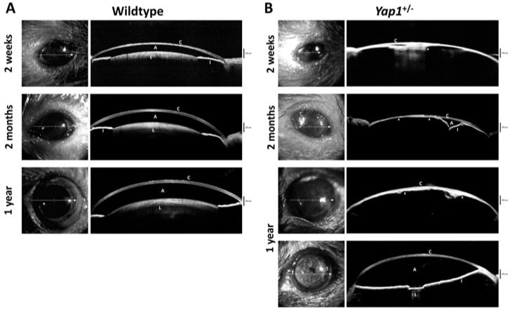
Complete ophthalmic examinations, as well as corneal esthesiometry, the phenol red thread test, intraocular pressure, and Fourier-domain optical coherence tomography were performed on and age-matched wild-type (WT) mice between eyelid opening (2 weeks after birth) and adulthood (2 months and 1 year after birth). Following euthanasia, enucleated eyes were characterized histologically.

RESULTS

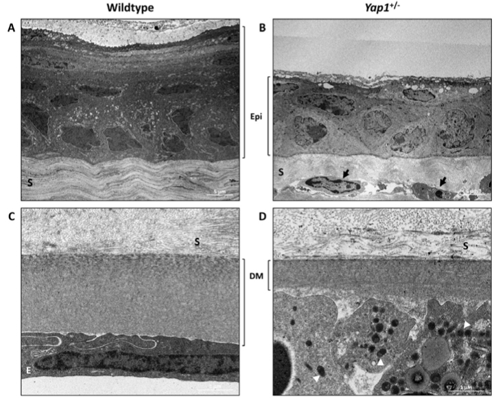
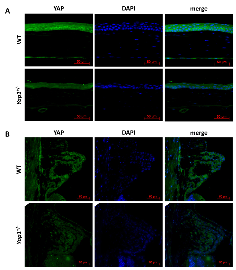
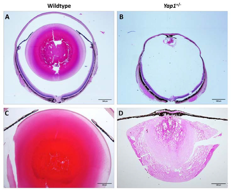
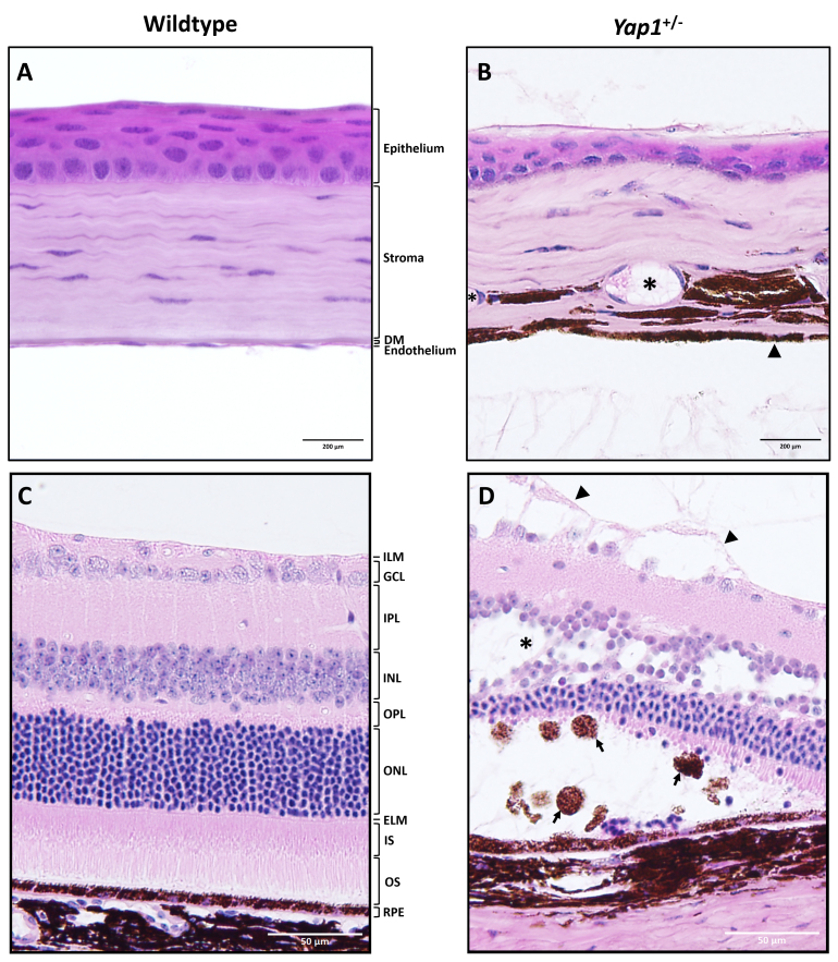
Microphthalmia with small palpebral fissures, corneal fibrosis, and reduced corneal sensation were common findings in the mice. Generalized corneal fibrosis precluded clinical examination of the posterior structures. Histologically, thinning and keratinization of the corneal epithelium were observed in the mice in comparison with the WT mice. Distorted collagen fiber arrangement and hypercellularity of keratocytes were observed in the stroma. Descemet's membrane was extremely thin and lacked an endothelial layer in the mice. The iris was adherent to the posterior cornea along most of its surface creating a distorted contour. Most of the eyes were microphakic with swollen fibers and bladder cells. The retinas of the mice were normal at 2 weeks and 2 months of age, but the presence of retinal abnormalities, including retinoschisis and detachment, was markedly increased in the mice at 1 year of age.

CONCLUSIONS

The results show that the heterozygous deletion of the gene in mice leads to complex ocular abnormalities, including microphthalmia, corneal fibrosis, anterior segment dysgenesis, and cataract.

摘要

目的

为了确定小鼠眼中( )单拷贝缺失的影响,详细测定了( )的眼部表型后果。

方法

对出生后睁眼(出生后2周)至成年期(出生后2个月和1年)的( )小鼠和年龄匹配的野生型(WT)小鼠进行了全面的眼科检查,以及角膜感觉测量、酚红棉线试验、眼压测量和傅里叶域光学相干断层扫描。安乐死后,对摘除的眼球进行组织学特征分析。

结果

小眼症伴睑裂小、角膜纤维化和角膜感觉减退是( )小鼠的常见表现。广泛性角膜纤维化妨碍了对后部结构的临床检查。组织学上,与WT小鼠相比,( )小鼠角膜上皮变薄并角化。基质中观察到胶原纤维排列紊乱和角膜细胞增多。( )小鼠的Descemet膜极薄且缺乏内皮细胞层。虹膜大部分表面与角膜后部粘连,轮廓扭曲。大多数( )小鼠眼球晶状体小,纤维和囊状细胞肿胀。( )小鼠在2周龄和2月龄时视网膜正常,但在1岁时,( )小鼠视网膜异常(包括视网膜劈裂和脱离)的发生率明显增加。

结论

结果表明,小鼠中该基因的杂合缺失导致复杂的眼部异常,包括小眼症、角膜纤维化、眼前节发育异常和白内障。

https://cdn.ncbi.nlm.nih.gov/pmc/blobs/1d29/6382475/2ac217413e27/mv-v25-129-f6.jpg
https://cdn.ncbi.nlm.nih.gov/pmc/blobs/1d29/6382475/b45d72218f7c/mv-v25-129-f1.jpg
https://cdn.ncbi.nlm.nih.gov/pmc/blobs/1d29/6382475/c554e93ae4a3/mv-v25-129-f2.jpg
https://cdn.ncbi.nlm.nih.gov/pmc/blobs/1d29/6382475/2ed6de15ad0e/mv-v25-129-f3.jpg
https://cdn.ncbi.nlm.nih.gov/pmc/blobs/1d29/6382475/77615f2e7ddb/mv-v25-129-f4.jpg
https://cdn.ncbi.nlm.nih.gov/pmc/blobs/1d29/6382475/c26e65ccbaf6/mv-v25-129-f5.jpg
https://cdn.ncbi.nlm.nih.gov/pmc/blobs/1d29/6382475/2ac217413e27/mv-v25-129-f6.jpg

相似文献

1
Ocular phenotypic consequences of a single copy deletion of the gene ( ) in mice.小鼠中该基因单拷贝缺失的眼部表型后果。 (括号内内容原文缺失,无法完整翻译)
Mol Vis. 2019 Feb 17;25:129-142. eCollection 2019.
2
Intracorneal positioning of the lens in Pax6-GAL4/VP16 transgenic mice.Pax6-GAL4/VP16转基因小鼠晶状体的角膜内定位
Mol Vis. 2005 Oct 26;11:876-86.
3
Histopathological characterisation of effects of the mouse Pax6(Leca4) missense mutation on eye development.小鼠Pax6(Leca4)错义突变对眼睛发育影响的组织病理学特征
Exp Eye Res. 2009 Aug;89(2):263-73. doi: 10.1016/j.exer.2009.03.016. Epub 2009 Apr 2.
4
A novel histopathologic finding in the Descemet's membrane of a patient with Peters Anomaly: a case-report and literature review.彼得斯异常患者后弹力层的一种新的组织病理学发现:病例报告及文献综述
BMC Ophthalmol. 2015 Oct 23;15:139. doi: 10.1186/s12886-015-0131-y.
5
Clinical and ultrastructural features of a novel hereditary anterior segment dysgenesis.一种新型遗传性眼前节发育异常的临床及超微结构特征
Ophthalmology. 2002 Mar;109(3):513-9. doi: 10.1016/s0161-6420(01)00975-7.
6
Ultrastructure of the posterior corneal stroma.后角膜基质的超微结构。
Ophthalmology. 2015 Apr;122(4):693-9. doi: 10.1016/j.ophtha.2014.09.037. Epub 2014 Nov 18.
7
Enlargement of the globe with ocular malformations in c-Myc transgenic mice.c-Myc转基因小鼠中眼球增大伴眼部畸形
Jpn J Ophthalmol. 1999 May-Jun;43(3):201-8. doi: 10.1016/s0021-5155(99)00014-3.
8
A Population Study of Common Ocular Abnormalities in C57BL/6N rd8 Mice.C57BL/6N rd8 小鼠常见眼部异常的群体研究。
Invest Ophthalmol Vis Sci. 2018 May 1;59(6):2252-2261. doi: 10.1167/iovs.17-23513.
9
Heterozygous loss-of-function mutations in YAP1 cause both isolated and syndromic optic fissure closure defects.YAP1 杂合性功能丧失突变可导致孤立型和综合征型视神经裂闭合缺陷。
Am J Hum Genet. 2014 Feb 6;94(2):295-302. doi: 10.1016/j.ajhg.2014.01.001. Epub 2014 Jan 23.
10
The requirement of pax6 for postnatal eye development: evidence from experimental mouse chimeras.出生后眼睛发育中Pax6的需求:来自实验性小鼠嵌合体的证据。
Invest Ophthalmol Vis Sci. 2007 Jul;48(7):3292-300. doi: 10.1167/iovs.06-1482.

引用本文的文献

1
The Hippo signalling pathway and its impact on eye diseases.Hippo 信号通路及其对眼部疾病的影响。
J Cell Mol Med. 2024 Apr;28(8):e18300. doi: 10.1111/jcmm.18300.
2
Uveitic glaucoma-like features in Yap conditional knockout mice.雅浦岛条件性基因敲除小鼠中的葡萄膜炎性青光眼样特征
Cell Death Discov. 2024 Jan 25;10(1):48. doi: 10.1038/s41420-023-01791-6.
3
Squishy matters - Corneal mechanobiology in health and disease.黏弹物质——健康与疾病中的角膜生物力学。

本文引用的文献

1
A Population Study of Common Ocular Abnormalities in C57BL/6N rd8 Mice.C57BL/6N rd8 小鼠常见眼部异常的群体研究。
Invest Ophthalmol Vis Sci. 2018 May 1;59(6):2252-2261. doi: 10.1167/iovs.17-23513.
2
Recapitulating X-Linked Juvenile Retinoschisis in Mouse Model by Knock-In Patient-Specific Novel Mutation.通过敲入患者特异性新突变在小鼠模型中重现X连锁青少年视网膜劈裂症
Front Mol Neurosci. 2018 Jan 12;10:453. doi: 10.3389/fnmol.2017.00453. eCollection 2017.
3
Retinal Degeneration Triggers the Activation of YAP/TEAD in Reactive Müller Cells.
Prog Retin Eye Res. 2024 Mar;99:101234. doi: 10.1016/j.preteyeres.2023.101234. Epub 2024 Jan 2.
4
Eyes open on stem cells.干细胞睁开了眼睛。
Stem Cell Reports. 2023 Dec 12;18(12):2313-2327. doi: 10.1016/j.stemcr.2023.10.023. Epub 2023 Nov 30.
5
The biophysical property of the limbal niche maintains stemness through YAP.角膜缘龛的生物物理特性通过 YAP 维持干细胞特性。
Cell Death Differ. 2023 Jun;30(6):1601-1614. doi: 10.1038/s41418-023-01156-7. Epub 2023 Apr 24.
6
Mice Deficient in TAZ (Wwtr1) Demonstrate Clinical Features of Late-Onset Fuchs' Endothelial Corneal Dystrophy.TAZ(WWTR1)缺陷小鼠表现出迟发性 Fuchs 内皮角膜营养不良的临床特征。
Invest Ophthalmol Vis Sci. 2023 Apr 3;64(4):22. doi: 10.1167/iovs.64.4.22.
7
CELF1 Selectively Regulates Alternative Splicing of DNA Repair Genes Associated With Cataract in Human Lens Cell Line.CELF1 选择性调节与人类晶状体细胞系白内障相关的 DNA 修复基因的可变剪接。
Biochem Genet. 2023 Aug;61(4):1319-1333. doi: 10.1007/s10528-022-10324-2. Epub 2022 Dec 30.
8
ARHGAP35 is a novel factor disrupted in human developmental eye phenotypes.ARHGAP35 是一种在人类发育性眼部表型中发生突变的新型因子。
Eur J Hum Genet. 2023 Mar;31(3):363-367. doi: 10.1038/s41431-022-01246-z. Epub 2022 Dec 1.
9
Animal models of corneal endothelial dysfunction to facilitate development of novel therapies.角膜内皮功能障碍的动物模型,以促进新型疗法的开发。
Ann Transl Med. 2021 Aug;9(15):1271. doi: 10.21037/atm-20-4389.
10
Genome-wide meta-analysis identifies 127 open-angle glaucoma loci with consistent effect across ancestries.全基因组荟萃分析确定了 127 个开角型青光眼基因座,这些基因座在不同种族中具有一致的效应。
Nat Commun. 2021 Feb 24;12(1):1258. doi: 10.1038/s41467-020-20851-4.
视网膜变性触发反应性穆勒细胞中YAP/TEAD的激活。
Invest Ophthalmol Vis Sci. 2017 Apr 1;58(4):1941-1953. doi: 10.1167/iovs.16-21366.
4
Yap is essential for retinal progenitor cell cycle progression and RPE cell fate acquisition in the developing mouse eye.Yap对于发育中小鼠眼睛的视网膜祖细胞周期进程和视网膜色素上皮(RPE)细胞命运获得至关重要。
Dev Biol. 2016 Nov 15;419(2):336-347. doi: 10.1016/j.ydbio.2016.09.001. Epub 2016 Sep 9.
5
Intrinsic and extrinsic regulatory mechanisms are required to form and maintain a lens of the correct size and shape.形成并维持大小和形状正确的晶状体需要内在和外在调节机制。
Exp Eye Res. 2017 Mar;156:34-40. doi: 10.1016/j.exer.2016.04.009. Epub 2016 Apr 21.
6
Yap1 Regulates Multiple Steps of Chondrocyte Differentiation during Skeletal Development and Bone Repair.Yap1在骨骼发育和骨修复过程中调控软骨细胞分化的多个步骤。
Cell Rep. 2016 Mar 8;14(9):2224-2237. doi: 10.1016/j.celrep.2016.02.021. Epub 2016 Feb 25.
7
Yap and Taz play a crucial role in neural crest-derived craniofacial development.Yes相关蛋白(Yap)和转录共激活因子(Taz)在神经嵴来源的颅面发育中发挥着关键作用。
Development. 2016 Feb 1;143(3):504-15. doi: 10.1242/dev.126920. Epub 2015 Dec 30.
8
Chronic inflammation imposes aberrant cell fate in regenerating epithelia through mechanotransduction.慢性炎症通过机械转导在再生上皮中施加异常的细胞命运。
Nat Cell Biol. 2016 Feb;18(2):168-80. doi: 10.1038/ncb3290. Epub 2015 Dec 21.
9
YAP controls retinal stem cell DNA replication timing and genomic stability.YAP控制视网膜干细胞的DNA复制时间和基因组稳定性。
Elife. 2015 Sep 22;4:e08488. doi: 10.7554/eLife.08488.
10
Yap and Taz regulate retinal pigment epithelial cell fate.Yes相关蛋白和转录共激活因子Taz调控视网膜色素上皮细胞命运。
Development. 2015 Sep 1;142(17):3021-32. doi: 10.1242/dev.119008. Epub 2015 Jul 24.