Department of Diagnostic and Interventional Radiology, Klinikum rechts der Isar, Technische Universitaet Muenchen, Munich, Germany.
Department of Nuclear Medicine, Klinikum rechts der Isar, Technische Universitaet Muenchen, Munich, Germany.
PLoS One. 2019 Mar 1;14(3):e0213082. doi: 10.1371/journal.pone.0213082. eCollection 2019.
To investigate whether signal to noise (SNR) analysis of contrast-enhanced MRI gives additional benefit for early disease detection by Magnetic Resonance Imaging (MRI) of experimental rheumatoid arthritis (RA) in a small animal model.
We applied contrast-enhanced MRI at 7T in DBA mice with or without collagen-induced arthritis (CIA). Clinical score, OMERACT RAMRIS analysis and analysis of signal to noise ratios (SNR) of regions of interest in RA bearing mice, methotrexate/methylprednisolone acetate treated RA and control animals were compared with respect to benefit for early diagnosis.
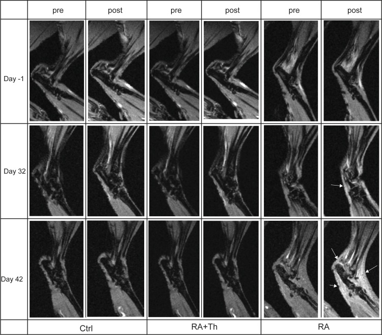
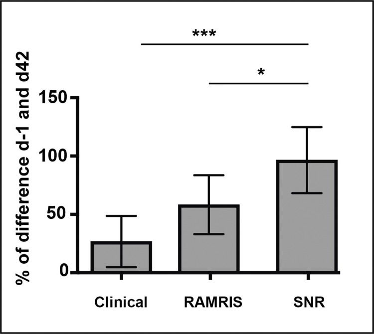
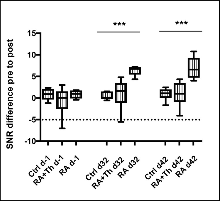
While treated RA and control animals did not show signs of RA activity in any of the above-mentioned scoring methods at any time point analyzed, RA animals revealed characteristic signs of RA in RAMRIS at the same time point when RA was detected clinically through scoring of the paws. The MR-based SNR analysis detected signs of synovitis, the earliest indication of RA, not only in late clinical stages, but also at an early stage when little or no clinical signs of RA were present in CIA animals and RAMRIS did not allow a distinct early detection.
SNR analysis of contrast-enhanced MR imaging provides additional benefit for early arthritis detection in CIA mice.
通过磁共振成像(MRI)对实验性类风湿关节炎(RA)的小动物模型进行研究,探讨信号噪声比(SNR)分析是否对早期疾病检测有额外的益处。
我们在患有或不患有胶原诱导关节炎(CIA)的 DBA 小鼠中应用了 7T 对比增强 MRI。临床评分、OMERACT RAMRIS 分析以及关节炎小鼠、甲氨蝶呤/醋酸甲基强的松龙治疗的关节炎和对照动物的感兴趣区域的 SNR 分析,在早期诊断方面进行了比较。
虽然治疗的 RA 和对照动物在任何分析的时间点都没有在任何上述评分方法中显示 RA 活动的迹象,但在通过爪子评分临床检测到 RA 的同时,RA 动物在 RAMRIS 中显示出 RA 的特征性迹象。基于 MR 的 SNR 分析不仅在晚期临床阶段,而且在 CIA 动物中出现少量或没有明显 RA 临床症状的早期阶段,也检测到滑膜炎的迹象,这是 RA 的最早迹象,而 RAMRIS 无法明确早期检测到滑膜炎。
对比增强磁共振成像的 SNR 分析为 CIA 小鼠的早期关节炎检测提供了额外的益处。