• 文献检索
  • 文档翻译
  • 深度研究
  • 学术资讯
  • Suppr Zotero 插件Zotero 插件
  • 邀请有礼
  • 套餐&价格
  • 历史记录
应用&插件
Suppr Zotero 插件Zotero 插件浏览器插件Mac 客户端Windows 客户端微信小程序
定价
高级版会员购买积分包购买API积分包
服务
文献检索文档翻译深度研究API 文档MCP 服务
关于我们
关于 Suppr公司介绍联系我们用户协议隐私条款
关注我们

Suppr 超能文献

核心技术专利:CN118964589B侵权必究
粤ICP备2023148730 号-1Suppr @ 2026

文献检索

告别复杂PubMed语法,用中文像聊天一样搜索,搜遍4000万医学文献。AI智能推荐,让科研检索更轻松。

立即免费搜索

文件翻译

保留排版,准确专业,支持PDF/Word/PPT等文件格式,支持 12+语言互译。

免费翻译文档

深度研究

AI帮你快速写综述,25分钟生成高质量综述,智能提取关键信息,辅助科研写作。

立即免费体验

高分辨率扩散加权成像结合长回波链读出分段技术在子宫内膜癌肌层浸润中的应用。

High resolution diffusion-weighted imaging with readout segmentation of long variable echo-trains for determining myometrial invasion in endometrial carcinoma.

机构信息

Department of Obstetrics and Gynecology, The Second Xiangya Hospital, Central South University, Changsha, No.139 Renmin Road, Changsha, Hunan, 410011, PR China.

Department of Radiology, The Second Xiangya Hospital, Central South University, Changsha, Hunan, PR China.

出版信息

Cancer Imaging. 2020 Sep 21;20(1):66. doi: 10.1186/s40644-020-00346-7.

DOI:10.1186/s40644-020-00346-7
PMID:32958041
原文链接:https://pmc.ncbi.nlm.nih.gov/articles/PMC7507745/
Abstract

BACKGROUND

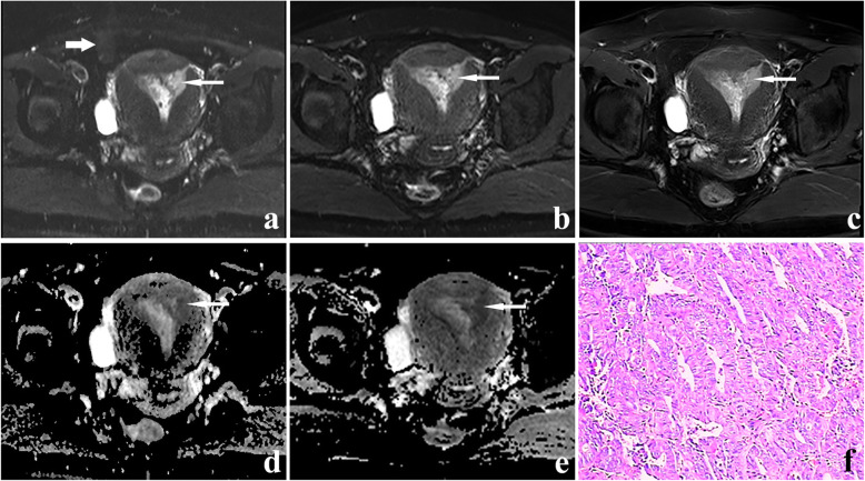
We assessed the image quality of endometrial cancer lesions by readout segmentation of long variable echo-trains (RESOLVE) diffusion-weighted imaging (DWI) compared with that by single-shot echo-planar imaging (SS-EPI) DWI, aimed to explore the value of RESOLVE DWI for determining myometrial invasion and clinical stage in endometrial cancer.

MATERIALS AND METHODS

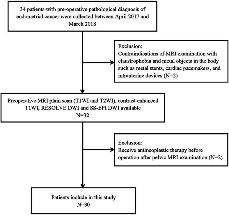
From April 2017 to March 2018, a total of 30 endometrial cancer patients (mean age 52.8 ± 9.0 years), who had undergone RESOLVE DWI and SS-EPI DWI, were included in the study. The image quality of endometrial carcinoma by two kinds of DWI scanning methods was compared qualitatively and quantitatively. The Spearman rank correlation test was used to assess the correlation of qualitative image quality scores between two readers. The accuracy of two DWI methods in detecting myometrial invasion and staging of endometrial carcinoma was calculated according to postoperative pathological results. The indexes were analyzed including sensitivity, specificity, accuracy, positive predictive value (PPV), and negative predictive value (NPV).

RESULTS

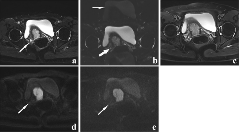
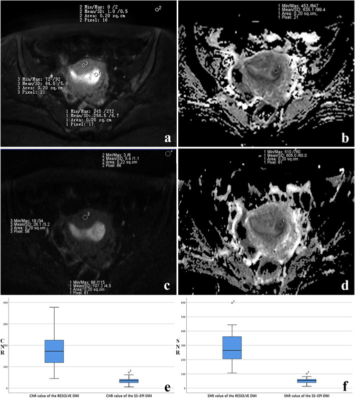
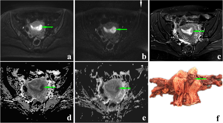
The qualitative score of RESOLVE DWI group was superior to SS-EPI DWI group in every aspect of five aspects (all P < 0.001). Interobserver agreement of depiction was good or excellent in two DWI sequences. Signal to noise ratio and contrast to noise ratio values in RESOLVE DWI group were both higher than those in SS-EPI DWI group (P<0.001). No statistical difference of apparent diffusion coefficient value was observed between two DWI groups (P = 0.261). The specificity, accuracy, PPV, and NPV of estimating myometrial invasion by RESOLVE DWI in three cases (intramucosal lesion, <50% superficial invasion and ≥ 50% deep invasion) were all higher than those by SS-EPI DWI for endometrial carcinoma. Especially RESOLVE DWI was valuable in judging <50% superficial invasion (95%CI:0.586, 0.970). No significant difference in accuracy staging was between the two DWI groups (P = 0.125).

CONCLUSION

RESOLVE DWI can provide higher quality images of endometrial carcinoma than SS-EPI DWI. The high-quality images are helpful for precise assessment of myometrial invasion in endometrial cancer.

摘要

背景

我们通过读取分段长变量回波序列(RESOLVE)弥散加权成像(DWI)与单次激发回波平面成像(SS-EPI)DWI 评估子宫内膜癌病变的图像质量,旨在探索 RESOLVE DWI 对确定子宫内膜癌肌层浸润和临床分期的价值。

材料与方法

2017 年 4 月至 2018 年 3 月,共纳入 30 例子宫内膜癌患者(平均年龄 52.8±9.0 岁),均行 RESOLVE DWI 和 SS-EPI DWI 检查。对两种 DWI 扫描方法对子宫内膜癌的图像质量进行定性和定量比较。采用 Spearman 秩相关检验评估两位观察者之间定性图像质量评分的相关性。根据术后病理结果计算两种 DWI 方法在检测子宫内膜癌肌层浸润和分期中的准确性。分析的指标包括灵敏度、特异度、准确性、阳性预测值(PPV)和阴性预测值(NPV)。

结果

在五个方面,RESOLVE DWI 组的定性评分均优于 SS-EPI DWI 组(均 P<0.001)。两种 DWI 序列的观察者间评估具有良好或极好的一致性。RESOLVE DWI 组的信噪比和对比噪声比均高于 SS-EPI DWI 组(均 P<0.001)。两种 DWI 组的表观弥散系数值无统计学差异(P=0.261)。RESOLVE DWI 对三种情况(黏膜内病变、<50%浅层浸润和≥50%深层浸润)估计子宫内膜癌肌层浸润的特异性、准确性、PPV 和 NPV 均高于 SS-EPI DWI。特别是 RESOLVE DWI 对<50%浅层浸润的评估有价值(95%CI:0.586,0.970)。两种 DWI 组的分期准确性无显著差异(P=0.125)。

结论

RESOLVE DWI 可提供比 SS-EPI DWI 更高质量的子宫内膜癌图像。高质量图像有助于精确评估子宫内膜癌的肌层浸润。

https://cdn.ncbi.nlm.nih.gov/pmc/blobs/fdf6/7507745/3c279ab253bd/40644_2020_346_Fig5_HTML.jpg
https://cdn.ncbi.nlm.nih.gov/pmc/blobs/fdf6/7507745/c24dbf24f5ea/40644_2020_346_Fig1_HTML.jpg
https://cdn.ncbi.nlm.nih.gov/pmc/blobs/fdf6/7507745/f3b52199743a/40644_2020_346_Fig2_HTML.jpg
https://cdn.ncbi.nlm.nih.gov/pmc/blobs/fdf6/7507745/57af53586538/40644_2020_346_Fig3_HTML.jpg
https://cdn.ncbi.nlm.nih.gov/pmc/blobs/fdf6/7507745/a7cfae032355/40644_2020_346_Fig4_HTML.jpg
https://cdn.ncbi.nlm.nih.gov/pmc/blobs/fdf6/7507745/3c279ab253bd/40644_2020_346_Fig5_HTML.jpg

相似文献

1
High resolution diffusion-weighted imaging with readout segmentation of long variable echo-trains for determining myometrial invasion in endometrial carcinoma.高分辨率扩散加权成像结合长回波链读出分段技术在子宫内膜癌肌层浸润中的应用。
Cancer Imaging. 2020 Sep 21;20(1):66. doi: 10.1186/s40644-020-00346-7.
2
Bladder cancer: detection and image quality compared among iShim, RESOLVE, and ss-EPI diffusion-weighted MR imaging with high b value at 3.0 T MRI.膀胱癌:在3.0T磁共振成像中,比较iShim、RESOLVE和具有高b值的单次激发自旋回波扩散加权磁共振成像在膀胱癌检测及图像质量方面的表现。
Medicine (Baltimore). 2017 Dec;96(50):e9292. doi: 10.1097/MD.0000000000009292.
3
Comparison of ZOOMit-DWI sequence and conventional DWI sequence in endometrial cancer.ZOOMit-DWI 序列与常规 DWI 序列在子宫内膜癌中的对比。
Zhong Nan Da Xue Xue Bao Yi Xue Ban. 2023 Jan 28;48(1):76-83. doi: 10.11817/j.issn.1672-7347.2023.220018.
4
Evaluation of multiplexed sensitivity encoding diffusion-weighted imaging in detecting uterine lesions: Image quality optimization.评价多重敏感编码扩散加权成像在检测子宫病变中的应用:图像质量优化。
Magn Reson Imaging. 2024 Jul;110:17-22. doi: 10.1016/j.mri.2024.03.003. Epub 2024 Mar 5.
5
Clinical comparison of single-shot EPI, readout-segmented EPI and TGSE-BLADE for diffusion-weighted imaging of cerebellopontine angle tumors on 3 tesla.3.0T 磁共振单次激发 EPI、分段读出 EPI 及 TGSE-BLADE 序列在桥小脑角肿瘤扩散加权成像中的临床对比研究。
Magn Reson Imaging. 2021 Dec;84:76-83. doi: 10.1016/j.mri.2021.09.009. Epub 2021 Sep 21.
6
Readout-segmented echo-planar imaging in the evaluation of sinonasal lesions: A comprehensive comparison of image quality in single-shot echo-planar imaging.读出分段回波平面成像在鼻窦病变评估中的应用:单次激发回波平面成像图像质量的全面比较
Magn Reson Imaging. 2016 Feb;34(2):166-72. doi: 10.1016/j.mri.2015.10.010. Epub 2015 Nov 2.
7
Preoperative Evaluation of Myometrial Invasion in Endometrial Carcinoma: Prospective Intra-individual Comparison of Magnetic Resonance Volumetry, Diffusion-weighted and Dynamic Contrast-enhanced Magnetic Resonance Imaging.子宫内膜癌肌层浸润的术前评估:磁共振容积测定法、扩散加权及动态对比增强磁共振成像的前瞻性个体内比较
Anticancer Res. 2018 Aug;38(8):4813-4817. doi: 10.21873/anticanres.12791.
8
Readout-segmented echo-planar imaging for diffusion-weighted imaging in the pelvis at 3T-A feasibility study.3T下盆腔扩散加权成像的读出分段回波平面成像——一项可行性研究
Acad Radiol. 2014 Apr;21(4):531-7. doi: 10.1016/j.acra.2014.01.005.
9
Readout-segmented echo-planar diffusion-weighted imaging in the assessment of orbital tumors: comparison with conventional single-shot echo-planar imaging in image quality and diagnostic performance.读出分段回波平面扩散加权成像在眼眶肿瘤评估中的应用:与传统单次激发回波平面成像在图像质量和诊断性能方面的比较。
Acta Radiol. 2017 Dec;58(12):1457-1467. doi: 10.1177/0284185117695667. Epub 2017 Mar 22.
10
Preoperative prediction of deep myometrial invasion and tumor grade for stage I endometrioid adenocarcinoma: a simple method of measurement on DWI.术前预测 I 期子宫内膜样腺癌的深肌层浸润和肿瘤分级:DWI 上的一种简单测量方法。
Eur Radiol. 2019 Feb;29(2):838-848. doi: 10.1007/s00330-018-5653-2. Epub 2018 Jul 23.

引用本文的文献

1
Deep learning reconstruction of diffusion-weighted imaging with single-shot echo-planar imaging in endometrial cancer: a comparison with multi-shot echo-planar imaging.子宫内膜癌中基于单次激发回波平面成像的扩散加权成像深度学习重建:与多次激发回波平面成像的比较
Abdom Radiol (NY). 2025 Apr 18. doi: 10.1007/s00261-025-04955-3.
2
Evaluating the image quality and local tumor invasion of uterine cancer by MUSE DWI with RPG.利用MUSE DWI结合RPG评估子宫癌的图像质量和局部肿瘤浸润情况。
Heliyon. 2024 Jul 30;10(15):e35440. doi: 10.1016/j.heliyon.2024.e35440. eCollection 2024 Aug 15.
3
Multi-b-values-fitting readout-segmentation of long variable echo-trains diffusion-weighted imaging (RESOLVE DWI) in evaluation of disease activity and curative effect of axial spondyloarthritis (axSpA).

本文引用的文献

1
Multi-b-value diffusion weighted imaging for preoperative evaluation of risk stratification in early-stage endometrial cancer.多 b 值弥散加权成像在早期子宫内膜癌术前风险分层评估中的应用。
Eur J Radiol. 2019 Oct;119:108637. doi: 10.1016/j.ejrad.2019.08.006. Epub 2019 Aug 12.
2
The added value of diffusion-weighted imaging in the preoperative assessment of endometrial cancer.扩散加权成像在子宫内膜癌术前评估中的附加价值。
Radiol Bras. 2019 Jul-Aug;52(4):229-236. doi: 10.1590/0100-3984.2018.0054.
3
Differential diagnosis of nasopharyngeal carcinoma and nasopharyngeal lymphoma based on DCE-MRI and RESOLVE-DWI.
多 b 值拟合读出分段长回波链扩散加权成像(RESOLVE DWI)在评价中轴型脊柱关节炎(axSpA)的疾病活动度和疗效中的应用。
Front Immunol. 2023 Jul 3;14:1136925. doi: 10.3389/fimmu.2023.1136925. eCollection 2023.
4
Image quality evaluation of diffusion-weighted imaging in bladder cancer: a comparison between integrated slice-specific dynamic shimming and single-shot echo-planar imaging.膀胱癌扩散加权成像的图像质量评估:集成切片特异性动态匀场与单次激发回波平面成像的比较
Quant Imaging Med Surg. 2023 Apr 1;13(4):2526-2537. doi: 10.21037/qims-22-851. Epub 2023 Feb 17.
5
Comparison of ZOOMit-DWI sequence and conventional DWI sequence in endometrial cancer.ZOOMit-DWI 序列与常规 DWI 序列在子宫内膜癌中的对比。
Zhong Nan Da Xue Xue Bao Yi Xue Ban. 2023 Jan 28;48(1):76-83. doi: 10.11817/j.issn.1672-7347.2023.220018.
6
Utility of the Diffusion Weighted Sequence in Gynecological Imaging: Review Article.扩散加权序列在妇科成像中的应用:综述文章。
Cancers (Basel). 2022 Sep 15;14(18):4468. doi: 10.3390/cancers14184468.
7
Apparent diffusion coefficient values in Modic changes - interobserver reproducibility and relation to Modic type.MRI 下 Modic 改变的表观弥散系数值:观察者间可重复性及其与 Modic 类型的关系。
BMC Musculoskelet Disord. 2022 Jul 22;23(1):695. doi: 10.1186/s12891-022-05610-4.
8
Role of MRI-Based Functional Imaging in Improving the Therapeutic Index of Radiotherapy in Cancer Treatment.基于磁共振成像的功能成像在提高癌症放疗治疗指数中的作用。
Front Oncol. 2021 Aug 27;11:645177. doi: 10.3389/fonc.2021.645177. eCollection 2021.
9
Diagnostic accuracy of high -value diffusion weighted imaging for patients with prostate cancer: a diagnostic comprehensive analysis.高价值弥散加权成像对前列腺癌患者的诊断准确性:一项诊断综合分析。
Aging (Albany NY). 2021 Jun 22;13(12):16404-16424. doi: 10.18632/aging.203164.
基于 DCE-MRI 和 RESOLVE-DWI 的鼻咽癌与鼻咽淋巴瘤的鉴别诊断。
Eur Radiol. 2020 Jan;30(1):110-118. doi: 10.1007/s00330-019-06343-0. Epub 2019 Aug 1.
4
SNR analysis of contrast-enhanced MR imaging for early detection of rheumatoid arthritis.对比增强磁共振成像的信噪比分析用于类风湿关节炎的早期检测。
PLoS One. 2019 Mar 1;14(3):e0213082. doi: 10.1371/journal.pone.0213082. eCollection 2019.
5
Comparative analysis of the value of diffusion kurtosis imaging and diffusion-weighted imaging in evaluating the histological features of endometrial cancer.扩散峰度成像与扩散加权成像评估子宫内膜癌组织学特征的价值比较分析。
Cancer Imaging. 2019 Feb 14;19(1):9. doi: 10.1186/s40644-019-0196-6.
6
Are ADC values of readout-segmented echo-planar diffusion-weighted imaging (RESOLVE) correlated with pathological prognostic factors in rectal adenocarcinoma?读片分段回波平面扩散加权成像(RESOLVE)的 ADC 值与直肠腺癌的病理预后因素相关吗?
World J Surg Oncol. 2018 Jul 12;16(1):138. doi: 10.1186/s12957-018-1445-z.
7
Inchworm sign of endometrial cancer on diffusion-weighted MRI: radiology-pathology correlation.子宫内膜癌在扩散加权磁共振成像上的尺蠖征:放射学与病理学相关性
Clin Radiol. 2018 Oct;73(10):907.e9-907.e14. doi: 10.1016/j.crad.2018.05.017. Epub 2018 Jun 9.
8
The relationship between addiction to smartphone usage and depression among adults: a cross sectional study.成年人智能手机使用成瘾与抑郁的关系:一项横断面研究。
BMC Psychiatry. 2018 May 25;18(1):148. doi: 10.1186/s12888-018-1745-4.
9
Differentiating Neuromyelitis Optica-Related and Multiple Sclerosis-Related Acute Optic Neuritis Using Conventional Magnetic Resonance Imaging Combined With Readout-Segmented Echo-Planar Diffusion-Weighted Imaging.使用传统磁共振成像结合读出分段回波平面扩散加权成像鉴别视神经脊髓炎相关和多发性硬化相关的急性视神经炎
J Comput Assist Tomogr. 2018 Jul/Aug;42(4):502-509. doi: 10.1097/RCT.0000000000000724.
10
Comparison of DWI Methods in the Pediatric Brain: PROPELLER Turbo Spin-Echo Imaging Versus Readout-Segmented Echo-Planar Imaging Versus Single-Shot Echo-Planar Imaging.儿童脑弥散加权成像方法的比较:螺旋桨涡轮自旋回波成像与分段读出回波平面成像与单次激发回波平面成像。
AJR Am J Roentgenol. 2018 Jun;210(6):1352-1358. doi: 10.2214/AJR.17.18796. Epub 2018 Mar 23.