Department of Anesthesiology, University of Colorado, Anschutz Medical Campus, Aurora, United States of America.
Department of Anesthesiology, University of Colorado, Anschutz Medical Campus, Aurora, United States of America; Neuroscience Graduate Program, University of Colorado, Anschutz Medical Campus, Aurora, United States of America.
Neurobiol Dis. 2019 Jul;127:472-481. doi: 10.1016/j.nbd.2019.01.016. Epub 2019 Feb 28.
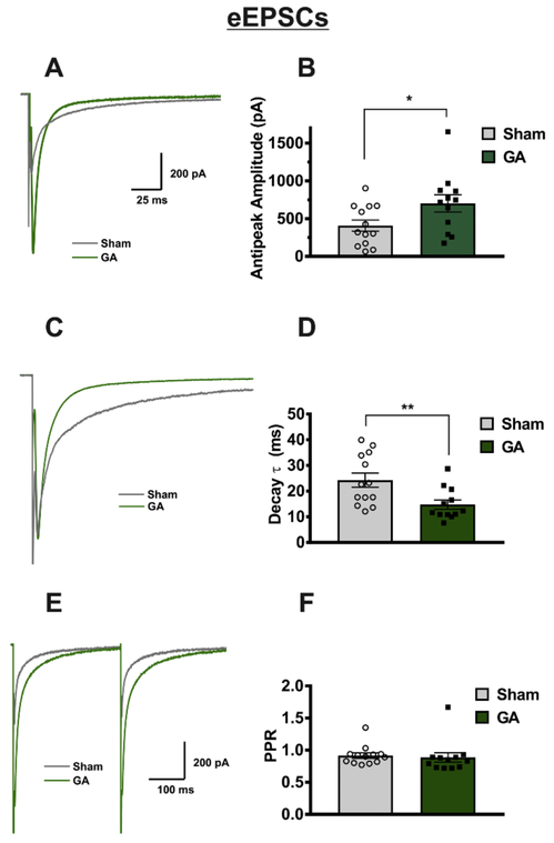
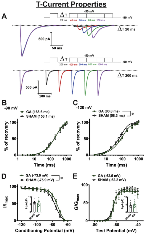
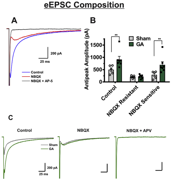
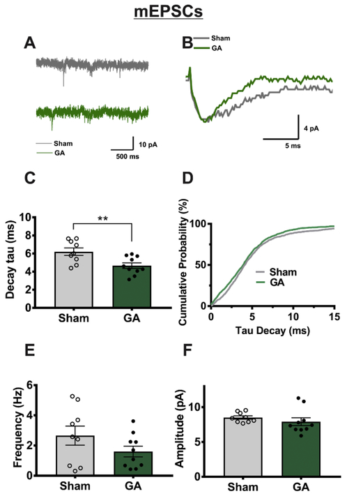
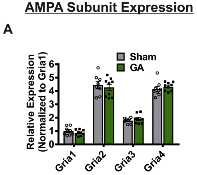
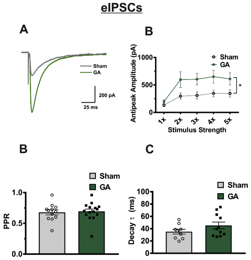
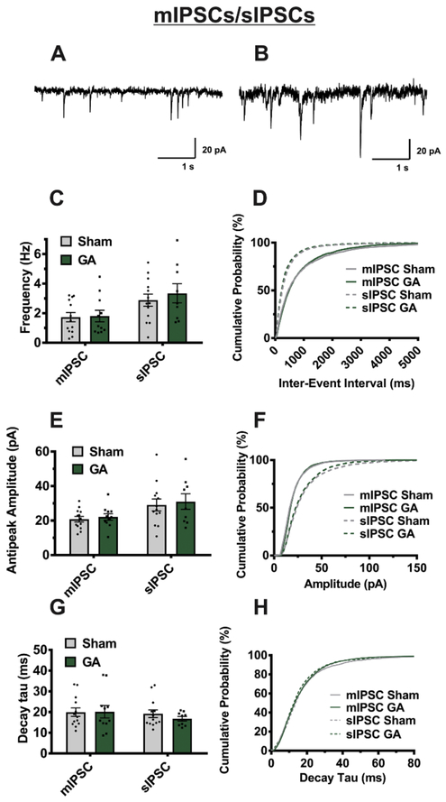
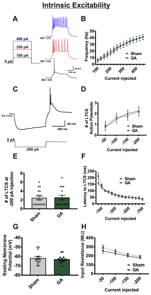
Ample evidence has surfaced documenting the neurotoxic effects of various general anesthetic (GA) agents in the mammalian brain when administered at critical periods of synaptogenesis. However, little is known about how this neurotoxic insult affects persisting neuronal excitability after the initial exposure. Here we investigated synaptic activity and intrinsic excitability of the ventrobasal nucleus (VB) of the thalamus caused by neonatal GA administration. We used patch-clamp recordings from acute thalamic slices in young rats up to two weeks after neurotoxic GA exposure of isoflurane and nitrous oxide for 6 h at postnatal age of 7 (P7) days. We found that GA exposure at P7 increases evoked excitatory postsynaptic currents (eEPSCs) two fold by means through AMPA mediated mechanisms, while NMDA component was spared. In addition, miniature EPSCs showed a faster decay rate in neurons from GA treated animals when compared to sham controls. Likewise, we discovered that the amplitudes of evoked inhibitory postsynaptic currents (eIPSCs) were increased in VB neurons from GA animals about two-fold. Interestingly, these results were observed in female but not male rats. In contrast, intrinsic excitability and properties of T-type voltage gated calcium currents were minimally affected by GA exposure. Together, these data further the idea that GAs cause lasting alterations in synaptic transmission and neuronal excitability depending upon the placing and connectivity of neurons in the thalamus. Given that function of thalamocortical circuits critically depends on the delicate balance between excitation and inhibition, future development of therapies aimed at addressing consequences of altered excitability in the developing brain by GAs may be an attractive possibility.
大量证据表明,各种全身麻醉 (GA) 药物在哺乳动物大脑的突触发生关键期给药时会产生神经毒性作用。然而,对于这种神经毒性损伤如何影响初始暴露后持续的神经元兴奋性知之甚少。在这里,我们研究了新生 GA 给药后,幼年大鼠背侧基底核 (VB) 的突触活动和内在兴奋性。我们使用急性丘脑切片中的膜片钳记录,在新生后第 7 天 (P7) 接受 6 小时异氟烷和一氧化二氮神经毒性 GA 暴露后的年轻大鼠中进行。我们发现,GA 暴露在 P7 时通过 AMPA 介导的机制将诱发的兴奋性突触后电流 (eEPSC) 增加两倍,而 NMDA 成分则不受影响。此外,与假手术对照相比,GA 处理动物的神经元中小型 EPSC 的衰减速率更快。同样,我们发现 GA 动物 VB 神经元的诱发抑制性突触后电流 (eIPSC) 幅度增加了约两倍。有趣的是,这些结果仅在雌性大鼠中观察到,而在雄性大鼠中则没有观察到。相比之下,GA 暴露对 T 型电压门控钙电流的内在兴奋性和特性的影响最小。总之,这些数据进一步表明,GA 会根据丘脑神经元的位置和连接性,导致突触传递和神经元兴奋性的持久改变。鉴于丘脑皮质回路的功能严重依赖于兴奋和抑制之间的微妙平衡,因此针对 GA 改变发育中大脑兴奋性的后果开发治疗方法可能是一个有吸引力的选择。