Park Shin Ae, Sledge Dodd, Monahan Colleen, Bartoe Joshua T, Komáromy András M
Department of Small Animal Clinical Sciences, Veterinary Medical Center, College of Veterinary Medicine, Michigan State University, 736 Wilson Road, East Lansing, MI, 48824, USA.
Michigan State University Veterinary Diagnostic Laboratory, Lansing, MI, USA.
BMC Vet Res. 2019 Mar 4;15(1):75. doi: 10.1186/s12917-019-1812-1.
Open angle glaucoma is the only type of primary glaucoma reported in Beagles. This case report describes a primary angle-closure glaucoma in a Beagle and its diagnostic and prognostic relevance.
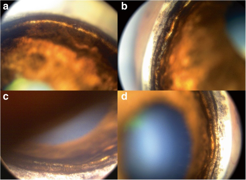
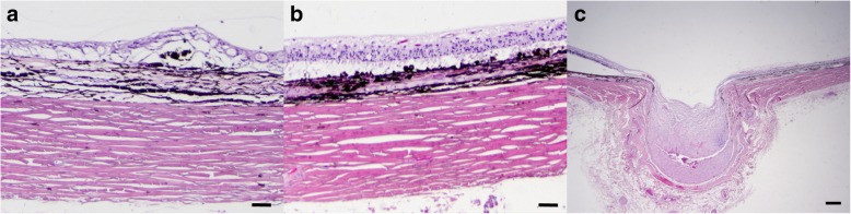
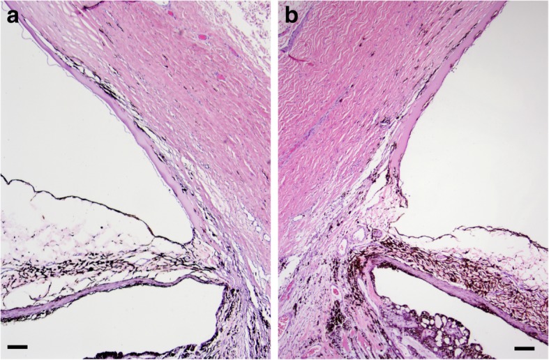
A 12-year-old, neutered male Beagle presented to the Michigan State University (MSU) Comparative Ophthalmology Service for evaluation of suspected visual impairment. Complete ophthalmic examination of the left eye (OS) revealed: blepharospasm, absent menace response, moderate episcleral congestion, mild diffuse corneal edema, mydriasis, asteroid hyalosis, decreased myelination and cupping of the optic nerve head, and mild retinal vascular attenuation. Examinations of the right eye (OD) were within normal limits. Intraocular Pressure (IOP) were 24 mmHg OD and 49 mmHg OS. Gonioscopy OD revealed a narrow iridocorneal angle with moderate pectinate ligament dysplasia characterized by broad-based pectinate ligament strands (fibrae latae) and solid sheets (laminae) throughout all 4 quadrants. DNA testing revealed that the dog did not carry the Gly661Arg ADAMTS10 mutation responsible for primary open angle glaucoma (POAG) in Beagles. The OS was medically managed with latanoprost 0.005% and dorzolamide HCl 2% /timolol malate 0.5% ophthalmic solutions for 7 months and then enucleated due to uncontrolled IOP. Histopathologic evaluation was consistent with goniodysgenesis with a broad, non-perforate, sheet-like band of uveal stroma bridging from the base of the iris to the terminal arborization of Descemet's membrane. Approximately 14 months from the initial diagnosis of glaucoma OS, OD also developed glaucoma and was enucleated. Histopathologic findings were consistent with goniodysgenesis OD.
To our knowledge, this is the first reported case of PACG with goniodysgenesis in a Beagle supported by clinical, genetic, and histopathologic data. It highlights the importance of gonioscopy in Beagles with glaucoma. Further studies with a larger number of dogs are warranted to characterize clinical manifestations and inheritance of PACG in this breed.
开角型青光眼是比格犬中报道的唯一一种原发性青光眼。本病例报告描述了一只比格犬的原发性闭角型青光眼及其诊断和预后相关性。
一只12岁已绝育的雄性比格犬因疑似视力受损就诊于密歇根州立大学(MSU)比较眼科服务中心。对左眼(OS)进行的全面眼科检查发现:眼睑痉挛、无威胁反应、中度巩膜表层充血、轻度弥漫性角膜水肿、瞳孔散大、星状玻璃体病变、视神经乳头髓鞘减少和杯状凹陷,以及轻度视网膜血管变细。右眼(OD)检查结果正常。眼压分别为右眼24 mmHg,左眼49 mmHg。右眼房角镜检查显示虹膜角膜角狭窄,伴有中度梳状韧带发育异常,其特征为在所有四个象限均有宽基底的梳状韧带束(纤维束)和实性薄片(板层)。DNA检测显示该犬不携带导致比格犬原发性开角型青光眼(POAG)的Gly661Arg ADAMTS10突变。左眼使用0.005%拉坦前列素和2%盐酸多佐胺/0.5%马来酸噻吗洛尔眼药水进行药物治疗7个月,随后因眼压控制不佳而被摘除眼球。组织病理学评估与房角发育异常一致,有一条宽阔的、无穿孔的、片状的葡萄膜基质带从虹膜基部延伸至Descemet膜的终末分支。在左眼青光眼初步诊断约14个月后,右眼也发生了青光眼并被摘除眼球。右眼组织病理学检查结果与房角发育异常一致。
据我们所知,这是第一例有临床、遗传和组织病理学数据支持的比格犬原发性闭角型青光眼伴房角发育异常的病例报告。它强调了房角镜检查在患有青光眼的比格犬中的重要性。有必要对更多的犬进行进一步研究,以明确该品种原发性闭角型青光眼的临床表现和遗传方式。