Department of Biomedical Engineering, College of Engineering and School of Medicine, Wayne State University, Detroit, MI, 48202, USA.
Positron Emission Tomography Center, Wayne State University, Detroit, MI, 48202, USA.
Sci Rep. 2019 Mar 5;9(1):3595. doi: 10.1038/s41598-019-40054-2.
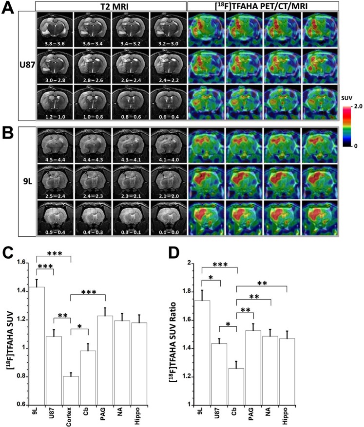
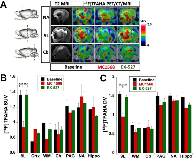
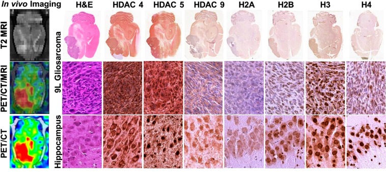
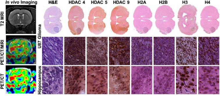
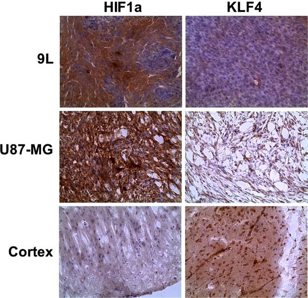
HDAC class IIa enzymes (HDAC4, 5, 7, 9) are important for glioma progression, invasion, responses to TMZ and radiotherapy, and prognosis. In this study, we demonstrated the efficacy of PET/CT/(MRI) with [F]TFAHA for non-invasive and quantitative imaging of HDAC class IIa expression-activity in intracerebral 9L and U87-MG gliomas in rats. Increased accumulation of [F]TFAHA in 9L and U87-MG tumors was observed at 20 min post radiotracer administration with SUV of 1.45 ± 0.05 and 1.08 ± 0.05, respectively, and tumor-to-cortex SUV ratios of 1.74 ± 0.07 and 1.44 ± 0.03, respectively. [F]TFAHA accumulation was also observed in normal brain structures known to overexpress HDACs class IIa: hippocampus, n.accumbens, PAG, and cerebellum. These results were confirmed by immunohistochemical staining of brain tissue sections revealing the upregulation of HDACs 4, 5, and 9, and HIF-1α, hypoacetylation of H2AK5ac, H2BK5ac, H3K9ac, H4K8ac, and downregulation of KLF4. Significant reduction in [F]TFAHA accumulation in 9L tumors was observed after administration of HDACs class IIa specific inhibitor MC1568, but not the SIRT1 specific inhibitor EX-527. Thus, PET/CT/(MRI) with [F]TFAHA can facilitate studies to elucidate the roles of HDAC class IIa enzymes in gliomagenesis and progression and to optimize therapeutic doses of novel HDACs class IIa inhibitors in gliomas.
组蛋白去乙酰化酶 IIa 类酶(HDAC4、5、7、9)在神经胶质瘤的进展、侵袭、对 TMZ 和放疗的反应以及预后中起着重要作用。在这项研究中,我们证明了 [F]TFAHA 的 PET/CT/(MRI)在大鼠脑内 9L 和 U87-MG 神经胶质瘤中对 HDAC Ⅱ a 表达-活性进行非侵入性和定量成像的有效性。在放射性示踪剂给药后 20 分钟,[F]TFAHA 在 9L 和 U87-MG 肿瘤中的积累增加,SUV 值分别为 1.45±0.05 和 1.08±0.05,肿瘤与皮质的 SUV 比值分别为 1.74±0.07 和 1.44±0.03。[F]TFAHA 的积累也观察到在已知过表达 HDACs Ⅱ a 的正常脑结构中:海马体、伏隔核、PAG 和小脑。这些结果通过脑组织切片的免疫组织化学染色得到证实,揭示了 HDACs 4、5 和 9 以及 HIF-1α 的上调,H2AK5ac、H2BK5ac、H3K9ac、H4K8ac 的低乙酰化和 KLF4 的下调。在给予 HDACs Ⅱ a 特异性抑制剂 MC1568 后,9L 肿瘤中 [F]TFAHA 的积累显著减少,但 SIRT1 特异性抑制剂 EX-527 则不然。因此,[F]TFAHA 的 PET/CT/(MRI)可以促进研究阐明 HDAC Ⅱ a 酶在神经胶质瘤发生和进展中的作用,并优化新型 HDAC Ⅱ a 抑制剂在神经胶质瘤中的治疗剂量。