Department of Neurology, University Hospital Bonn, Bonn, Germany.
Institute of Neuroscience and Medicine (INM-1), Research Center Juelich, Juelich, Germany.
PLoS One. 2019 Mar 7;14(3):e0213381. doi: 10.1371/journal.pone.0213381. eCollection 2019.
The aim of this study was to examine the natural history of brain involvement in adult-onset myotonic dystrophies type 1 and 2 (DM1, DM2).
We conducted a longitudinal observational study to examine functional and structural cerebral changes in myotonic dystrophies. We enrolled 16 adult-onset DM1 patients, 16 DM2 patients, and 17 controls. At baseline and after 5.5 ± 0.4 years participants underwent neurological, neuropsychological, and 3T-brain MRI examinations using identical study protocols that included voxel-based morphometry and diffusion tensor imaging. Data were analyzed by (i) group comparisons between patients and controls at baseline and follow-up, and (ii) group comparisons using difference maps (baseline-follow-up in each participant) to focus on disease-related effects over time.
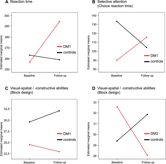
We found minor neuropsychological deficits with mild progression in DM1 more than DM2. Daytime sleepiness was restricted to DM1, whereas fatigue was present in both disease entities and stable over time. Comparing results of cross-sectional neuroimaging analyses at baseline and follow-up revealed an unchanged pattern of pronounced white matter alterations in DM1. There was mild additional gray matter reduction in DM1 at follow-up. In DM2, white matter reduction was of lesser extent, but there were some additional alterations at follow-up. Gray matter seemed unaffected in DM2. Intriguingly, longitudinal analyses using difference maps and comparing them between patients and controls did not reveal any significant differences of cerebral changes over time between patients and controls.
The lack of significant disease-related progression of gray and white matter involvement over a period of five years in our cohort of DM1 and DM2 patients suggests either a rather slowly progressive process or even a stable course of cerebral changes in middle-aged adult-onset patients. Being the first longitudinal neuroimaging trial in DM1 and DM2, this study provides useful additional information regarding the natural history of brain involvement.
本研究旨在探讨成年型肌强直性营养不良 1 型和 2 型(DM1、DM2)患者脑受累的自然病程。
我们进行了一项纵向观察性研究,以检测肌强直性营养不良患者的大脑功能和结构变化。共纳入 16 例成年起病的 DM1 患者、16 例 DM2 患者和 17 名对照者。在基线时和 5.5±0.4 年后,所有参与者均采用相同的研究方案进行神经学、神经心理学和 3T 脑 MRI 检查,包括基于体素的形态测量学和弥散张量成像。数据分析包括:(i)患者与对照组在基线和随访时的组间比较,以及(ii)采用差值图(每位参与者的基线-随访)进行组间比较,以关注疾病相关的随时间变化的效应。
我们发现 DM1 患者存在轻微的神经心理学缺陷,且随时间进展更为显著,而 DM2 患者则无进展。日间嗜睡仅限于 DM1,而疲劳在两种疾病实体中均存在且随时间稳定。比较基线和随访时的横断面神经影像学分析结果显示,DM1 的白质改变模式不变。DM1 患者在随访时出现轻微的额外灰质减少。在 DM2 中,白质减少程度较轻,但在随访时存在一些额外改变。DM2 患者的灰质似乎未受影响。有趣的是,使用差值图进行的纵向分析,并将其在患者和对照组之间进行比较,并未显示患者和对照组之间的脑变化在随时间推移有任何显著差异。
在我们的 DM1 和 DM2 患者队列中,经过五年的随访,未见灰质和白质受累有明显的与疾病相关的进展,这提示在中年起病的患者中,脑受累的过程可能较为缓慢,甚至呈稳定状态。作为 DM1 和 DM2 的首次纵向神经影像学研究,本研究提供了有关脑受累自然病程的有用信息。