Univ Rennes, CNRS, Inria, Inserm, IRISA UMR 6074, Empenn ERL U-1228, F-35000 Rennes, France.
Univ Rennes, CNRS, Inria, Inserm, IRISA UMR 6074, Empenn ERL U-1228, F-35000 Rennes, France; Centre Hospitalier Guillaume Régnier, Academic Psychiatry Department, 35703 Rennes, France; Univ of Rennes, "Behavior and Basal Ganglia" research unit (EA 4712), Rennes, France.
Neuroimage Clin. 2019;22:101710. doi: 10.1016/j.nicl.2019.101710. Epub 2019 Feb 4.
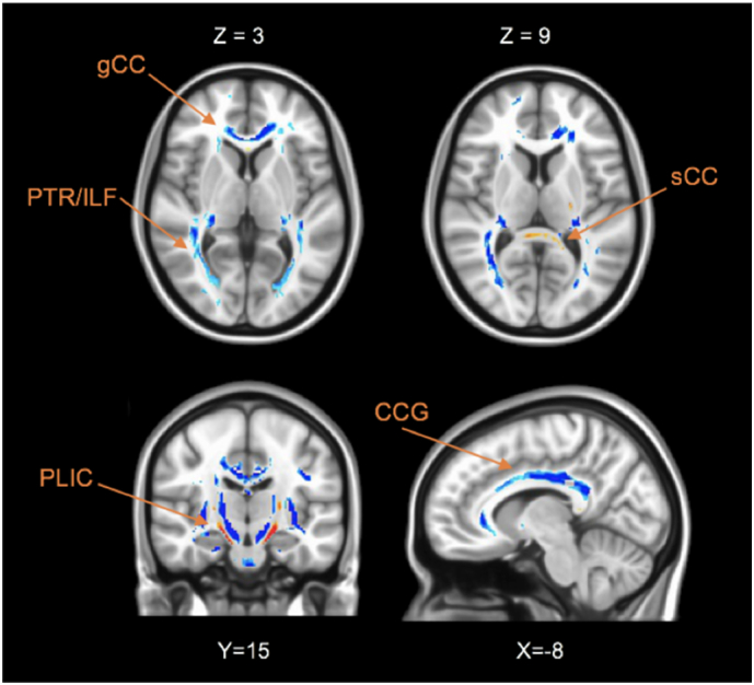
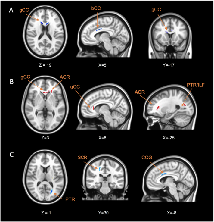
Mood depressive disorder is one of the most disabling chronic diseases with a high rate of everyday life disability that affects 350 million people around the world. Recent advances in neuroimaging have reported widespread structural abnormalities, suggesting a dysfunctional frontal-limbic circuit involved in the pathophysiological mechanisms of depression. However, a variety of different white matter regions has been implicated and is sought to suffer from lack of reproducibility of such categorical-based biomarkers. These inconsistent results might be attributed to various factors: actual categorical definition of depression as well as clinical phenotype variability. In this study, we 1/ examined WM changes in a large cohort (114 patients) compared to a healthy control group and 2/ sought to identify specific WM alterations in relation to specific depressive phenotypes such as anhedonia (i.e. lack of pleasure), anxiety and psychomotor retardation -three core symptoms involved in depression. Consistent with previous studies, reduced white matter was observed in the genu of the corpus callosum extending to the inferior fasciculus and posterior thalamic radiation, confirming a frontal-limbic circuit abnormality. Our analysis also reported other patterns of increased fractional anisotropy and axial diffusivity as well as decreased apparent diffusion coefficient and radial diffusivity in the splenium of the corpus callosum and posterior limb of the internal capsule. Moreover, a positive correlation between FA and anhedonia was found in the superior longitudinal fasciculus as well as a negative correlation in the cingulum. Then, the analysis of the anxiety and diffusion metric revealed that increased anxiety was associated with greater FA values in genu and splenium of corpus callosum, anterior corona radiata and posterior thalamic radiation. Finally, the motor retardation analysis showed a correlation between increased Widlöcher depressive retardation scale scores and reduced FA in the body and genu of the corpus callosum, fornix, and superior striatum. Through this twofold approach (categorical and phenotypic), this study has underlined the need to move forward to a symptom-based research area of biomarkers, which help to understand the pathophysiology of mood depressive disorders and to stratify precise phenotypes of depression with targeted therapeutic strategies.
心境性抑郁障碍是最具致残性的慢性疾病之一,全球有 3.5 亿人受到影响,日常生活功能障碍的发生率较高。神经影像学的最新进展报告了广泛的结构异常,表明存在涉及抑郁病理生理机制的额-边缘回路功能障碍。然而,各种不同的白质区域都受到牵连,并且由于基于分类的生物标志物的可重复性缺乏而受到关注。这些不一致的结果可能归因于以下各种因素:抑郁的实际分类定义以及临床表型的可变性。在这项研究中,我们 1/ 比较了 114 名患者和健康对照组的 WM 变化,2/ 试图确定与特定抑郁表型(如快感缺失,即缺乏愉悦感)、焦虑和精神运动迟滞有关的特定 WM 改变-抑郁的三个核心症状。与之前的研究一致,我们观察到胼胝体膝部的白质减少,延伸至下束和后丘脑辐射,证实了额-边缘回路异常。我们的分析还报告了其他模式的分数各向异性和轴向弥散度增加,以及胼胝体压部和内囊后肢的表观弥散系数和径向弥散度降低。此外,在胼胝体上纵束中发现 FA 与快感缺失呈正相关,在扣带中呈负相关。然后,对焦虑和扩散指标的分析表明,焦虑增加与胼胝体膝部和压部、前冠状辐射和后丘脑辐射的 FA 值增加相关。最后,运动迟滞分析显示,Widlöcher 抑郁迟滞量表评分的增加与胼胝体体部和膝部、穹窿和上纹状体的 FA 值降低相关。通过这种双重方法(分类和表型),本研究强调需要推进基于症状的生物标志物研究领域,这有助于了解心境性抑郁障碍的病理生理学,并针对具有靶向治疗策略的抑郁的精确表型进行分层。