Department of Electrical and Mechanical Engineering, Graduate School of Engineering, Nagoya Institute of Technology, Nagoya, Japan.
Department of Techno-Business Administration, Graduate School of Engineering, Nagoya Institute of Technology, Nagoya, Japan.
Sci Rep. 2019 Mar 8;9(1):3960. doi: 10.1038/s41598-019-40578-7.
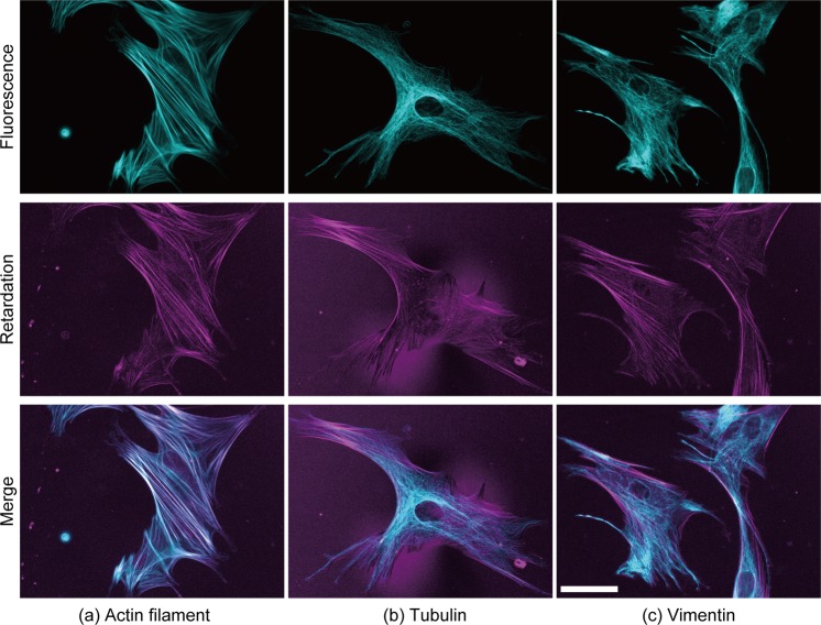
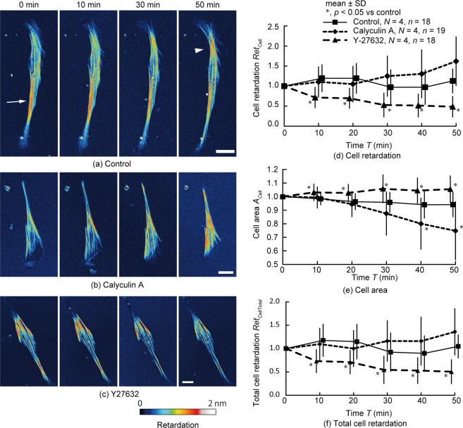
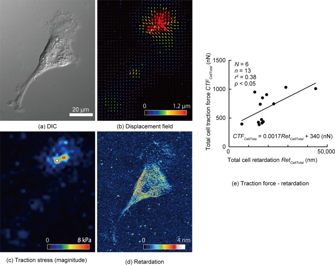
Vascular smooth muscle cells (VSMCs) have two distinct phenotypes: contractile and synthetic. The major difference between these phenotypes lies in the magnitude of the contractile force produced by the cell. Although traction force microscopy (TFM) is often used to evaluate cellular contractile force, this method requires complex preprocessing and a sufficiently compliant substrate. To evaluate the contractile force and the phenotype of living VSMCs with minimal effort and in a manner independent of the substrate stiffness, we propose a photoelasticity-based method using retardation, which is related to the difference between the first and second principal stresses and their orientation. The results demonstrate that actin filaments co-localize with areas of high retardation in cells, indicating that the retardation of VSMCs is promoted by actin filaments. The retardation of cells treated with calyculin A and Y-27632 tended to be larger and smaller, respectively, than that of control cells. Cell traction force significantly correlates with total cell retardation (r = 0.38). The retardation of contractile VSMCs (passage 2) was significantly higher than that of synthetic VSMCs (passage 12). These results indicate that cell retardation can be used to assess cell contractile force and, thus, determine the phenotype of VSMCs.
血管平滑肌细胞 (VSMCs) 具有两种截然不同的表型:收缩型和合成型。这两种表型的主要区别在于细胞产生的收缩力的大小。尽管牵引力量显微镜 (TFM) 常用于评估细胞的收缩力,但该方法需要复杂的预处理和足够的顺应性基底。为了在不依赖基底刚度的情况下以最小的努力评估活 VSMCs 的收缩力和表型,我们提出了一种基于光弹性的方法,使用延迟,延迟与第一和第二主应力及其方向之间的差异有关。结果表明,肌动蛋白丝与细胞中高延迟区域共定位,表明 VSMCs 的延迟是由肌动蛋白丝促进的。用 calyculin A 和 Y-27632 处理的细胞的延迟分别趋于大于和小于对照细胞。细胞牵引力与总细胞延迟显著相关 (r = 0.38)。收缩型 VSMCs (第 2 代) 的延迟明显高于合成型 VSMCs (第 12 代)。这些结果表明,细胞延迟可用于评估细胞收缩力,从而确定 VSMCs 的表型。