Xin Wenchong, Yang Xiaoyu, Wang Jianfeng, Shao Xiaoliang, Zhang Feifei, Shi Yunmei, Liu Bao, Yu Wenji, Tang Haipeng, Wu Zhifang, Wang Yuetao, Zhou Weihua
Departments of Nuclear Medicine.
Cardiology, The Third Affiliated Hospital of Soochow University, Changzhou, Jiangsu Province.
Nucl Med Commun. 2019 May;40(5):491-498. doi: 10.1097/MNM.0000000000001009.
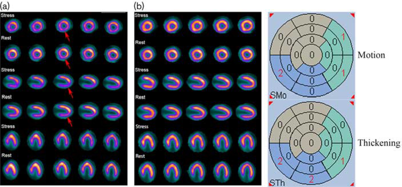
In single-photon emission computed tomography (SPECT) myocardial perfusion imaging (MPI) studies, attenuation artifacts frequently cause false positives, which can be partially overcome by computed tomography attenuation correction (CT-AC) or gated acquisition [gated myocardial perfusion imaging (GMPI)]. The purpose of this study is to evaluate their relative diagnostic performances for coronary artery disease (CAD).
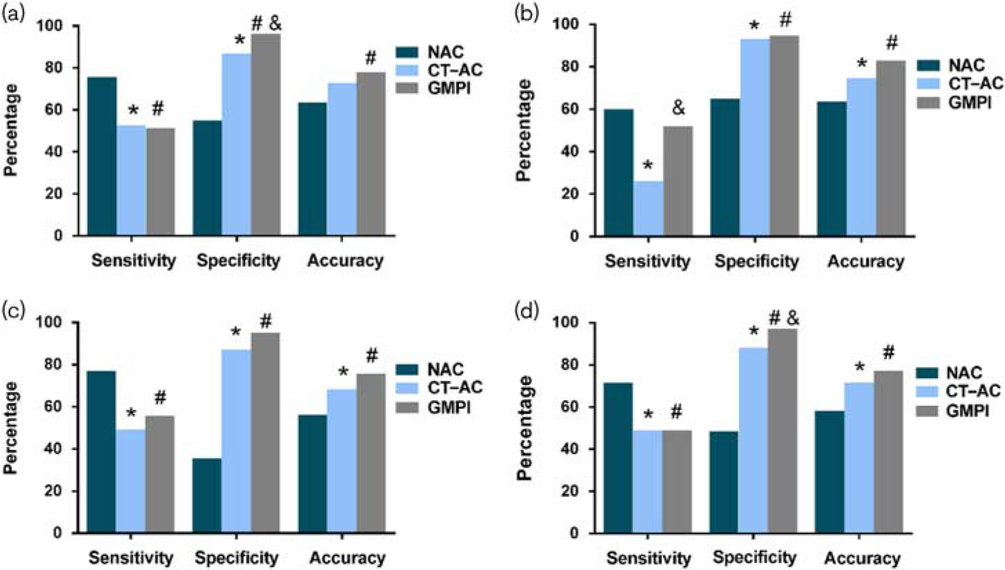
We enrolled 181 patients who underwent gated SPECT with CT-AC in this study. Two observers who were blinded to the clinical data interpreted the GMPI and CT-AC images. Coronary angiography was considered as the reference standard. The diagnostic efficacy was evaluated based on sex, BMI, and individual coronary arteries.
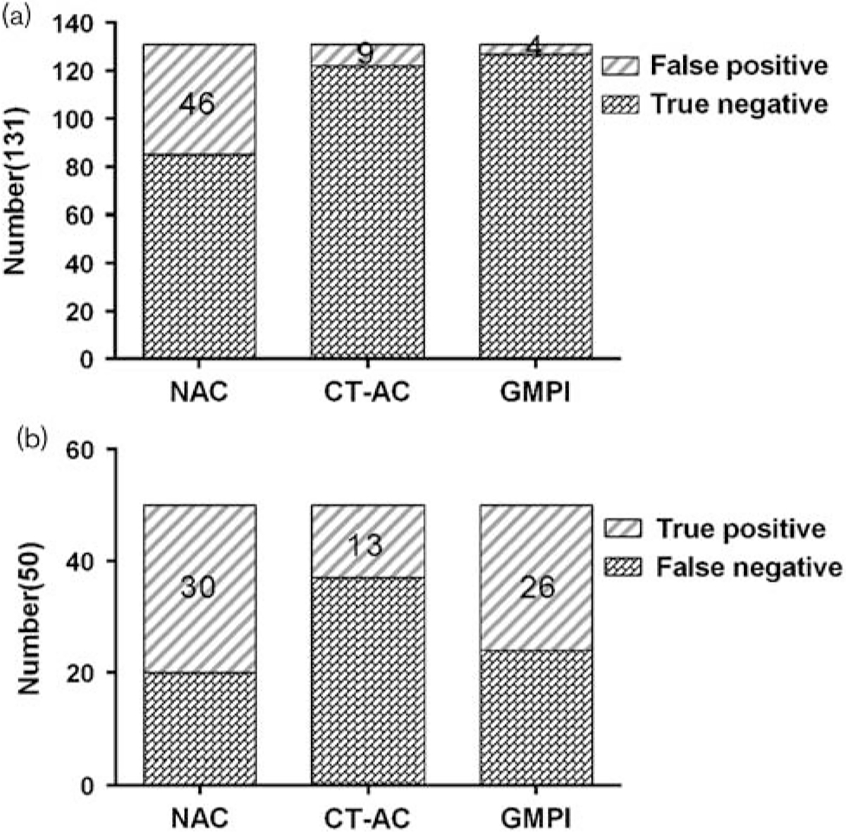
The diagnostic accuracy of GMPI was higher than that of nonattenuation correction overall, as well as for men, overweight individuals, and right CAD (P<0.05). Compared with CT-AC, GMPI overall had a higher specificity (96.3 vs. 86.9%, P=0.014) but the same sensitivity, achieving an increased accuracy and area under the curve (AUC, P>0.05). For diagnosing right CAD, GMPI had a higher diagnostic efficacy (AUC: 0.733 vs. 0.596, P<0.001) because of its higher sensitivity (52.0 vs. 26.0%, P=0.008); for men, the diagnostic efficacy of GMPI was significantly higher than that of CT-AC (AUC: 0.754 vs. 0.681, P=0.038).
Both CT-AC and GMPI led to an increased diagnostic efficacy compared with nonattenuation correction in differentiating attenuation artifacts from fixed perfusion defects. These improvements were, however, more obvious for GMPI than for CT-AC, especially in men and right CAD.
在单光子发射计算机断层扫描(SPECT)心肌灌注成像(MPI)研究中,衰减伪影经常导致假阳性结果,而计算机断层扫描衰减校正(CT-AC)或门控采集[门控心肌灌注成像(GMPI)]可部分克服这一问题。本研究旨在评估它们对冠状动脉疾病(CAD)的相对诊断性能。
本研究纳入了181例行CT-AC门控SPECT检查的患者。两名对临床资料不知情的观察者解读GMPI和CT-AC图像。冠状动脉造影被视为参考标准。根据性别、体重指数(BMI)和各支冠状动脉评估诊断效能。
总体而言,GMPI的诊断准确性高于未进行衰减校正时,在男性、超重个体和右冠状动脉CAD患者中也是如此(P<0.05)。与CT-AC相比,GMPI总体具有更高的特异性(96.3%对86.9%,P=0.014),但敏感性相同,准确性和曲线下面积(AUC)增加(P>0.05)。对于诊断右冠状动脉CAD,GMPI具有更高的诊断效能(AUC:0.733对0.596,P<0.001),因为其敏感性更高(52.0%对26.0%,P=0.008);对于男性,GMPI的诊断效能显著高于CT-AC(AUC:0.754对0.681,P=0.038)。
与未进行衰减校正相比,CT-AC和GMPI在区分衰减伪影与固定灌注缺损方面均提高了诊断效能。然而,这些改善在GMPI中比在CT-AC中更明显,尤其是在男性和右冠状动脉CAD患者中。