Division of Dental Anesthesiology, Niigata University Graduate School of Medical and Dental Sciences, Niigata, Japan.
Center for Advanced Oral Sciences, Niigata University Graduate School of Medical and Dental Sciences, Niigata, Japan.
Sci Rep. 2019 Mar 12;9(1):4245. doi: 10.1038/s41598-018-37819-6.
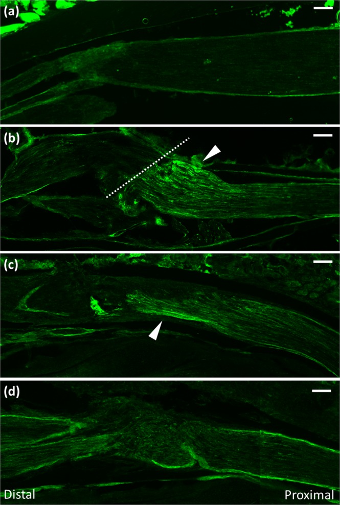
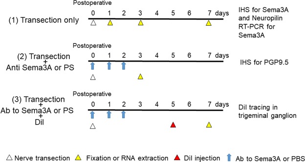
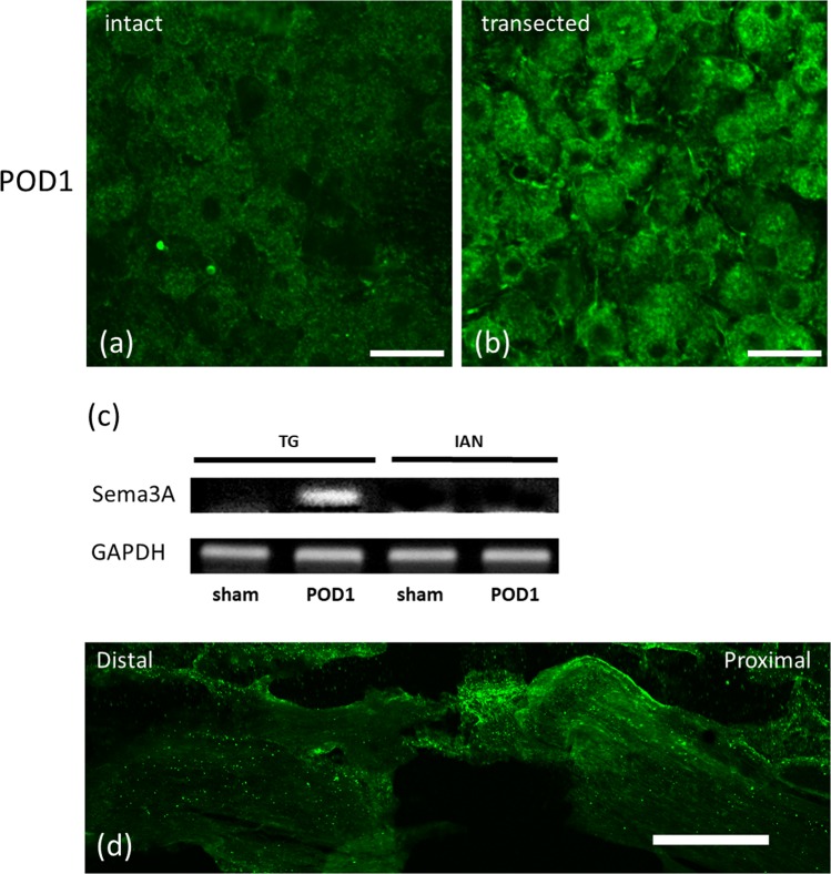
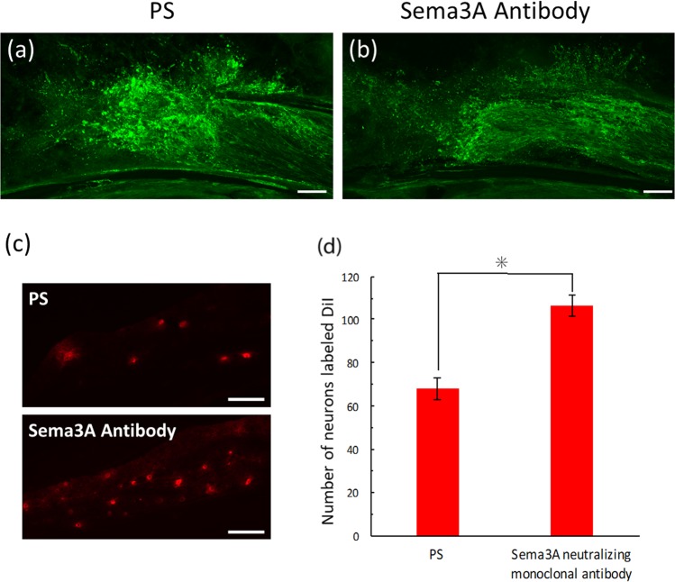
Neuroma formation at sites of injury can impair peripheral nerve regeneration. Although the involvement of semaphorin 3A has been suggested in neuroma formation, this detailed process after injury is not fully understood. This study was therefore undertaken to examine the effects of semaphorin 3A on peripheral nerve regeneration during the early stage after injury. Immunohistochemistry for semaphorin 3A and PGP9.5, a general neuronal marker, was carried out for clarify chronological changes in their expressions after transection of the mouse inferior alveolar nerve thorough postoperative days 1 to 7. At postoperative day 1, the proximal stump of the damaged IAN exhibited semaphorin 3A, while the distal stump lacked any immunoreactivity. From this day on, its expression lessened, ultimately disappearing completely in all regions of the transected inferior alveolar nerve. A local administration of an antibody to semaphorin 3A into the nerve transection site at postoperative day 3 inhibited axon sprouting at the injury site. This antibody injection increased the number of trigeminal ganglion neurons labeled with DiI (paired t-test, p < 0.05). Immunoreactivity of the semaphorin 3A receptor, neuropilin-1, was also detected at the proximal stump at postoperative day 1. These results suggest that nerve injury initiates semaphorin 3A production in ganglion neurons, which is then delivered through the nerve fibers to the proximal end, thereby contributes to the inhibition of axonal sprouting from the proximal region of injured nerves in the distal direction. To our knowledge, this is the first report to reveal the involvement of Sema3A in the nerve regeneration process at its early stage.
神经瘤的形成会损害周围神经的再生。尽管已经有人提出 Sema3A 在神经瘤形成中的作用,但损伤后这一详细过程尚未完全了解。因此,本研究旨在研究 Sema3A 在损伤后早期对外周神经再生的影响。通过对小鼠下颌下神经横断术后 1 至 7 天的免疫组织化学分析,研究 Sema3A 和 PGP9.5(一种通用神经元标记物)的表达变化。在术后第 1 天,损伤的 IAN 近端残端显示 Sema3A,而远端残端没有免疫反应性。从这一天开始,其表达减少,最终在所有横断的下颌下神经区域完全消失。术后第 3 天,在神经横断部位局部给予 Sema3A 抗体,可抑制损伤部位的轴突发芽。这种抗体注射增加了用 DiI 标记的三叉神经节神经元的数量(配对 t 检验,p < 0.05)。术后第 1 天,在近端残端还检测到 Sema3A 受体神经纤毛蛋白-1 的免疫反应性。这些结果表明,神经损伤会引发神经元中 Sema3A 的产生,然后通过神经纤维传递到近端,从而有助于抑制受损神经近端向远端的轴突发芽。据我们所知,这是首次报道 Sema3A 参与早期神经再生过程。