Faculty of Pharmacy, Kindai University, Higashi-Osaka 577-8502, Japan,
Faculty of Pharmacy, Keio University, Minato-ku, Tokyo 105-8512, Japan.
Int J Nanomedicine. 2019 Feb 18;14:1213-1227. doi: 10.2147/IJN.S196681. eCollection 2019.
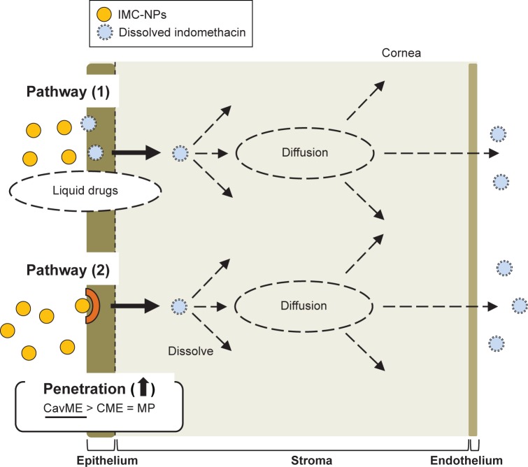
We previously found that ophthalmic formulations containing nanoparticles prepared by a bead mill method lead to an increase in bioavailability in comparison with traditional formulations (solution type). However, the transcorneal penetration pathway for ophthalmic formulations has not been explained yet. In this study, we investigated the mechanism of transcorneal penetration in the application of ophthalmic formulations containing indomethacin nanoparticles (IMC-NPs).
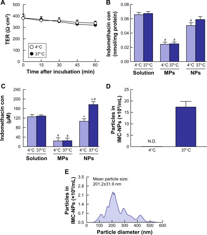
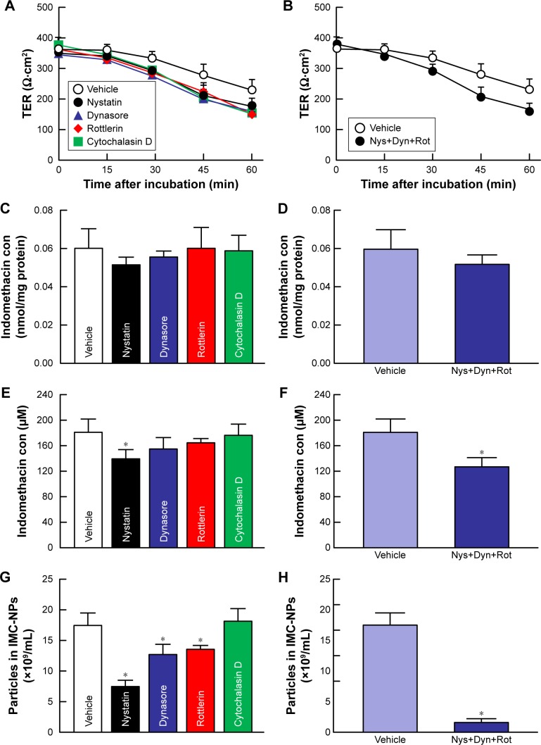
IMC-NPs was prepared by the bead mill method. For the analysis of energy-dependent endocytosis, corneal epithelial (HCE-T) cell monolayers and removed rabbit cornea were thermoregulated at 4°C, where energy-dependent endocytosis is inhibited. In addition, for the analysis of different endocytosis pathways using pharmacological inhibitors, inhibitors of caveolae-mediated endocytosis (54 µM nystatin), clathrin-mediated endocytosis (40 µM dynasore), macropinocytosis (2 µM rottlerin) or phagocytosis (10 µM cytochalasin D) were used.
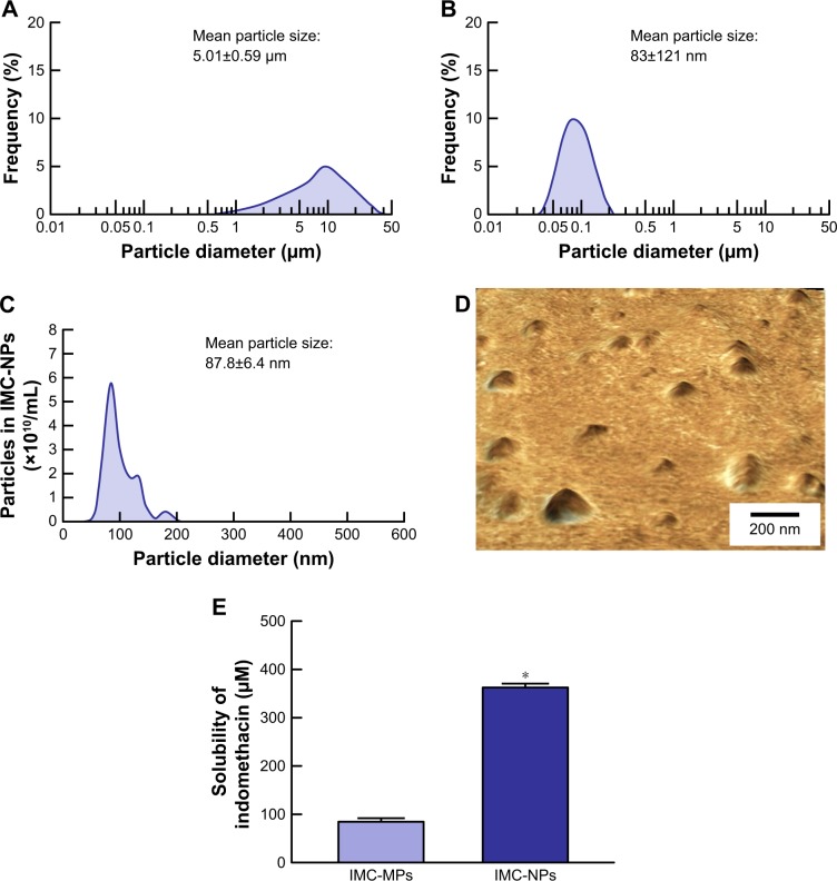
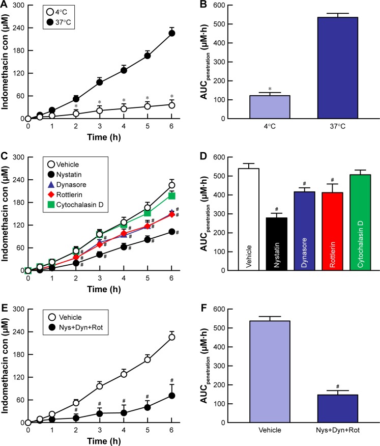
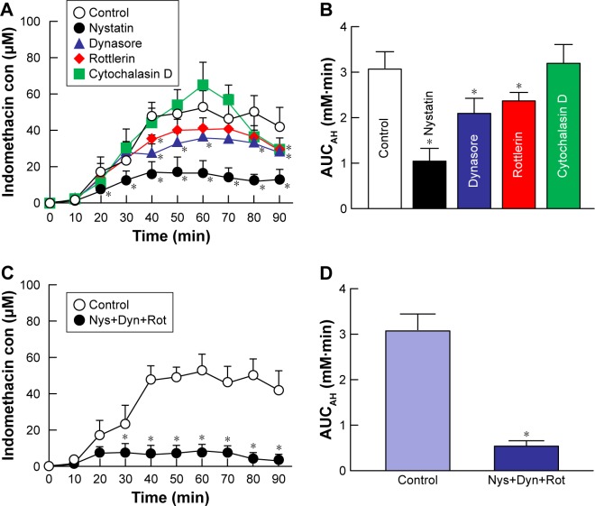
The ophthalmic formulations containing 35-200 nm sized indomethacin nanoparticles were prepared by treatment with a bead mill, and no aggregation or degradation of indomethacin was observed in IMC-NPs. The transcorneal penetration of indomethacin was significantly decreased by the combination of nystatin, dynasore and rottlerin, and the decreased penetration levels were similar to those at 4°C in HCE-T cell monolayers and rabbit cornea. In the in vivo experiments using rabbits, dynasore and rottlerin tended to decrease the transcorneal penetration of indomethacin (area under the drug concentration - time curve in the aqueous humor [AUC]), and the AUC in the nystatin-treated rabbit was significantly lower than that in non-treatment group. In addition, the AUC in rabbit corneas undergoing multi-treatment was obviously lower than that in rabbit corneas treated with each individual endocytosis inhibitor.
We found that three energy-dependent endocytosis pathways (clathrin-dependent endocytosis, caveolae-dependent endocytosis and macropinocytosis) are related to the trans-corneal penetration of indomethacin nanoparticles. In particular, the caveolae-dependent endocytosis is strongly involved.
我们之前发现,与传统制剂(溶液型)相比,通过珠磨机法制备的含纳米颗粒的眼科制剂可提高生物利用度。然而,眼科制剂的经角膜渗透途径尚未得到解释。在这项研究中,我们研究了含吲哚美辛纳米颗粒(IMC-NPs)的眼科制剂应用中的经角膜渗透机制。
通过珠磨机法制备 IMC-NPs。为了分析能量依赖性内吞作用,角膜上皮(HCE-T)细胞单层和去除的兔角膜在 4°C 下进行温度调节,在该温度下,能量依赖性内吞作用受到抑制。此外,为了使用药理学抑制剂分析不同的内吞途径,使用了 caveolae 介导的内吞作用抑制剂(54 µM 制霉菌素)、网格蛋白介导的内吞作用抑制剂(40 µM dynasore)、巨胞饮抑制剂(2 µM rottlerin)或吞噬作用抑制剂(10 µM 细胞松弛素 D)。
通过珠磨机处理制备了含有 35-200nm 大小的吲哚美辛纳米颗粒的眼科制剂,在 IMC-NPs 中未观察到吲哚美辛的聚集或降解。制霉菌素、dynasore 和 rottlerin 的联合使用显著降低了吲哚美辛的经角膜渗透,降低的渗透水平与 HCE-T 细胞单层和兔角膜中 4°C 时的水平相似。在使用兔的体内实验中,dynasore 和 rottlerin 倾向于降低吲哚美辛的经角膜渗透(房水中的药物浓度-时间曲线下面积[AUC]),并且制霉菌素处理的兔的 AUC 明显低于未处理组。此外,多处理兔角膜的 AUC 明显低于单独用每种内吞抑制剂处理的兔角膜。
我们发现三种能量依赖性内吞作用途径(网格蛋白依赖性内吞作用、小窝依赖性内吞作用和巨胞饮作用)与吲哚美辛纳米颗粒的经角膜渗透有关。特别是,小窝依赖性内吞作用强烈参与其中。