Huang Kui, Shen Lei, Niu Tieming, Zhao Ying, Fu Jiucun, Cao Yunpeng
Department of Neurology, The First Affiliated Hospital of China Medical University, Shenyang, Liaoning 110001, China.
Department of Geriatric Medicine, Shenyang Red Cross hospital, Shenyang Liaoning 110000, China.
Evid Based Complement Alternat Med. 2019 Feb 5;2019:2702068. doi: 10.1155/2019/2702068. eCollection 2019.
BACKGROUND/AIMS: Naomaitai can improve blood perfusion and ameliorate the damage in the paraventricular white matter. This study was focused on observing the neuroprotective effect of Naomaitai on the vascular dementia of rat and exploring the action mechanism of PI3K/PDK1/AKT signaling pathway.
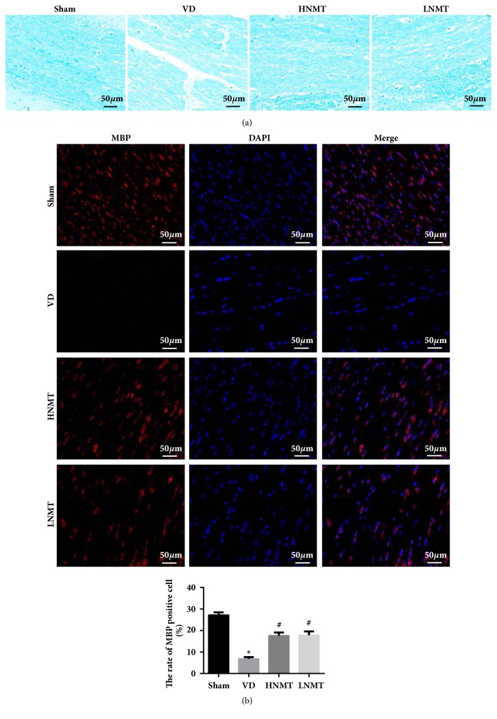
A vascular dementia model of rats was established by permanent, bilateral common carotid artery occlusion. Rats' behavior was tested by Neurological deficit score and the Morris water maze. The pathology and apoptosis were detected through HE staining and TUNEL assay. Myelin sheath loss and nerve fiber damage were detected by LFB staining. Inflammatory factors, oxidative stress, and brain damage markers were detected through ELISA. The expression of apoptosis-related proteins and PI3K/PDK1/AKT signaling pathway related proteins were measured by western blot. The expressions of PI3K, PDK1, AKT, and MBP in paraventricular white matter cells were detected by immunofluorescence.
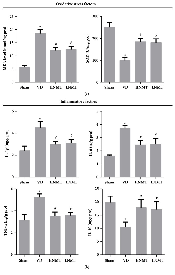
Naomaitai treatment decreased neurological function score in rats with vascular dementia, ameliorated paraventricular white matter damage caused by long-term hypoxia, and hypoperfusion reduced the brain injury markers S-100 and NSE contents, suppressed inflammatory reaction and oxidative stress, reduced IL-1, IL-6, TNF-, and MDA contents, and remarkably increased IL-10 and SOD contents. TUNEL and western blot assay showed that Naomaitai treatment decreased neuronal cell apoptosis, increased Bcl-2 expression, and reduced caspase-3 and Bax expression. Furthermore, we found Naomaitai inhibited PI3K and PDK1 expression and activated phosphorylated AKT protein in rats with vascular dementia. However, the protective effect of Naomatai in rats with vascular dementia was inhibited, and expression of PI3K signaling pathway-related proteins was blocked after administration of PI3K inhibitor.
Naomaitai can ameliorate brain damage in rats with vascular dementia, inhibit neuronal apoptosis, and have anti-inflammatory and antioxidative stress effects, which may be regulated by the PI3K/PDK1/AKT signaling pathway.
背景/目的:脑脉泰能改善血液灌注并减轻脑室旁白质损伤。本研究旨在观察脑脉泰对大鼠血管性痴呆的神经保护作用,并探讨PI3K/PDK1/AKT信号通路的作用机制。
通过永久性双侧颈总动脉闭塞建立大鼠血管性痴呆模型。采用神经功能缺损评分和莫里斯水迷宫测试大鼠行为。通过HE染色和TUNEL检测法检测病理学和细胞凋亡情况。通过LFB染色检测髓鞘脱失和神经纤维损伤。通过ELISA检测炎症因子、氧化应激和脑损伤标志物。通过蛋白质免疫印迹法检测凋亡相关蛋白和PI3K/PDK1/AKT信号通路相关蛋白的表达。通过免疫荧光检测脑室旁白质细胞中PI3K、PDK1、AKT和髓鞘碱性蛋白(MBP)的表达。
脑脉泰治疗降低了血管性痴呆大鼠的神经功能评分,改善了长期缺氧和灌注不足所致的脑室旁白质损伤,降低了脑损伤标志物S-100和神经元特异性烯醇化酶(NSE)的含量,抑制了炎症反应和氧化应激,降低了白细胞介素-1(IL-1)、白细胞介素-6(IL-6)、肿瘤坏死因子-α(TNF-α)和丙二醛(MDA)的含量,并显著提高了白细胞介素-10(IL-10)和超氧化物歧化酶(SOD)的含量。TUNEL和蛋白质免疫印迹分析表明,脑脉泰治疗减少了神经元细胞凋亡,增加了Bcl-2表达,并降低了半胱天冬酶-3(caspase-3)和Bax表达。此外,我们发现脑脉泰抑制血管性痴呆大鼠中PI3K和PDK1表达,并激活磷酸化AKT蛋白。然而,给予PI3K抑制剂后,脑脉泰对血管性痴呆大鼠的保护作用受到抑制,且PI3K信号通路相关蛋白的表达被阻断。
脑脉泰可改善血管性痴呆大鼠的脑损伤,抑制神经元凋亡,并具有抗炎和抗氧化应激作用,其作用可能受PI3K/PDK1/AKT信号通路调控。