Institute of Cancer Research, Radiotherapy and Imaging, London, United Kingdom.
Radiotherapy Department, Royal Marsden NHS Foundation Trust, London, United Kingdom.
Int J Radiat Oncol Biol Phys. 2019 Jul 1;104(3):685-693. doi: 10.1016/j.ijrobp.2019.03.003. Epub 2019 Mar 11.
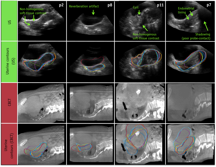
Adaptive radiation therapy strategies could account for interfractional uterine motion observed in patients with cervix cancer, but the current cone beam computed tomography (CBCT)-based treatment workflow is limited by poor soft-tissue contrast. The goal of the present study was to determine if ultrasound (US) could be used to improve visualization of the uterus, either as a single modality or in combination with CBCT.
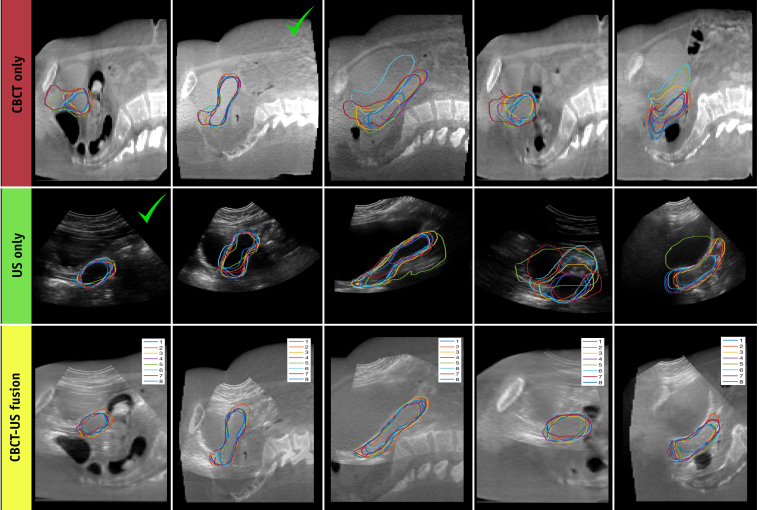
Interobserver uterine contour agreement and confidence were compared on 40 corresponding CBCT, US, and CBCT-US-fused images from 11 patients with cervix cancer. Contour agreement was measured using the Dice similarity coefficient (DSC) and mean contour-to-contour distance (MCCD). Observers rated their contour confidence on a scale from 1 to 10. Pairwise Wilcoxon signed-rank tests were used to measure differences in contour agreement and confidence.
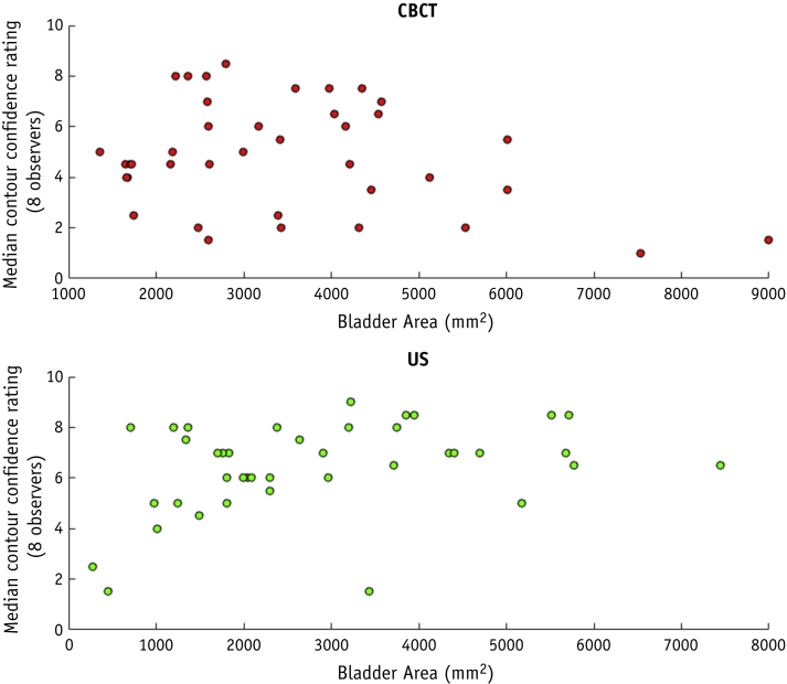
CBCT-US fused images had significantly better contour agreement and confidence than either individual modality (P < .05), with median (interquartile range [IQR]) values of 0.84 (0.11), 1.26 (0.23) mm, and 7 (2) for the DSC, MCCD, and observer confidence ratings, respectively. Contour agreement was similar between US and CBCT, with median (IQR) DSCs of 0.81 (0.17) and 0.82 (0.14) and MCCDs of 1.75 (1.15) mm and 1.62 (0.74) mm. Observers were significantly more confident in their US-based contours than in their CBCT-based contours (P < .05), with median (IQR) confidence ratings of 7 (2.75) versus 5 (4).
CBCT and US are complementary and improve uterine segmentation precision when combined. Observers could localize the uterus with a similar precision on independent US and CBCT images.
自适应放射治疗策略可以解释宫颈癌患者观察到的分次间子宫运动,但目前基于锥形束计算机断层扫描(CBCT)的治疗工作流程受到软组织对比度差的限制。本研究的目的是确定超声(US)是否可用于改善子宫的可视化,无论是作为单一模态还是与 CBCT 联合使用。
对 11 例宫颈癌患者的 40 对相应的 CBCT、US 和 CBCT-US 融合图像进行了观察者子宫轮廓的一致性和置信度比较。使用 Dice 相似系数(DSC)和平均轮廓到轮廓距离(MCCD)测量轮廓一致性。观察者对其轮廓置信度进行了 1 到 10 的评分。使用配对 Wilcoxon 符号秩检验来测量轮廓一致性和置信度的差异。
CBCT-US 融合图像的轮廓一致性和置信度明显优于任何单一模态(P<0.05),其 DSC、MCCD 和观察者置信度评分的中位数(四分位数范围[IQR])分别为 0.84(0.11)、1.26(0.23)mm 和 7(2)。US 和 CBCT 之间的轮廓一致性相似,其 DSCs 的中位数(IQR)分别为 0.81(0.17)和 0.82(0.14),MCCDs 的中位数(IQR)分别为 1.75(1.15)mm 和 1.62(0.74)mm。观察者对基于 US 的轮廓的置信度明显高于基于 CBCT 的轮廓(P<0.05),中位数(IQR)置信度评分为 7(2.75)与 5(4)。
CBCT 和 US 是互补的,联合使用可提高子宫分割精度。观察者可以在独立的 US 和 CBCT 图像上以相似的精度定位子宫。