• 文献检索
  • 文档翻译
  • 深度研究
  • 学术资讯
  • Suppr Zotero 插件Zotero 插件
  • 邀请有礼
  • 套餐&价格
  • 历史记录
应用&插件
Suppr Zotero 插件Zotero 插件浏览器插件Mac 客户端Windows 客户端微信小程序
定价
高级版会员购买积分包购买API积分包
服务
文献检索文档翻译深度研究API 文档MCP 服务
关于我们
关于 Suppr公司介绍联系我们用户协议隐私条款
关注我们

Suppr 超能文献

核心技术专利:CN118964589B侵权必究
粤ICP备2023148730 号-1Suppr @ 2026

文献检索

告别复杂PubMed语法,用中文像聊天一样搜索,搜遍4000万医学文献。AI智能推荐,让科研检索更轻松。

立即免费搜索

文件翻译

保留排版,准确专业,支持PDF/Word/PPT等文件格式,支持 12+语言互译。

免费翻译文档

深度研究

AI帮你快速写综述,25分钟生成高质量综述,智能提取关键信息,辅助科研写作。

立即免费体验

基于计算机断层扫描和磁共振成像的宫颈癌近距离放疗靶区勾画比较

Comparison of computed tomography- and magnetic resonance imaging-based target delineation for cervical cancer brachytherapy.

作者信息

Wang Fang, Bu Luyi, Wu Qun, Jiang Xue, Wu Lingyun, Li Yu, Xi Bin, Lu Zhongjie, Yan Senxiang

机构信息

Department of Radiotherapy, First Affiliated Hospital, Zhejiang University School of Medicine, Hangzhou, China.

Department of General Surgery, First Affiliated Hospital, Zhejiang University School of Medicine, Hangzhou, China.

出版信息

J Contemp Brachytherapy. 2020 Aug;12(4):367-374. doi: 10.5114/jcb.2020.98117. Epub 2020 Aug 21.

DOI:10.5114/jcb.2020.98117
PMID:33293976
原文链接:https://pmc.ncbi.nlm.nih.gov/articles/PMC7690230/
Abstract

PURPOSE

The objective of this study was to compare and assess the accuracy of computed tomography (CT)-based target delineation with that of magnetic resonance imaging (MRI)-based on high-dose-rate brachytherapy (HDR-BT) for patients with cervical cancer.

MATERIAL AND METHODS

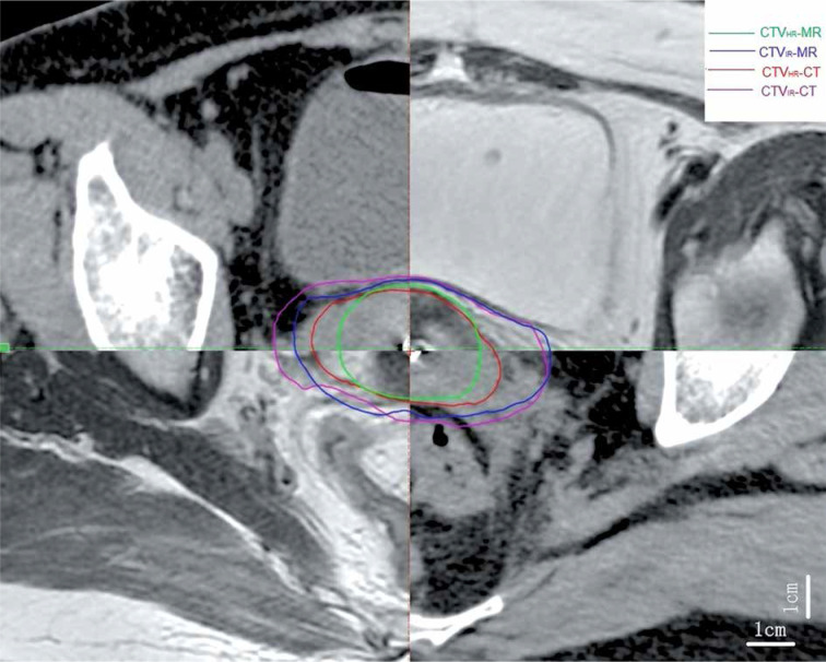
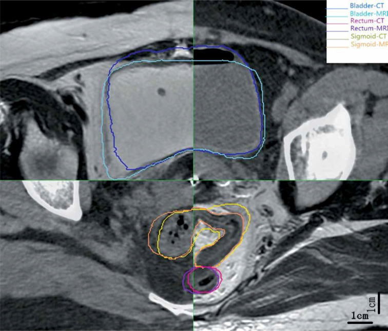
Data of 20 patients with locally advanced cervical cancer were collected and evaluated. Dimensions, conformity, and dose parameters of high-risk clinical target volume (CTV) as well as D, D, and D of organs at risk (OARs) based on MRI were compared with those based on CT.

RESULTS

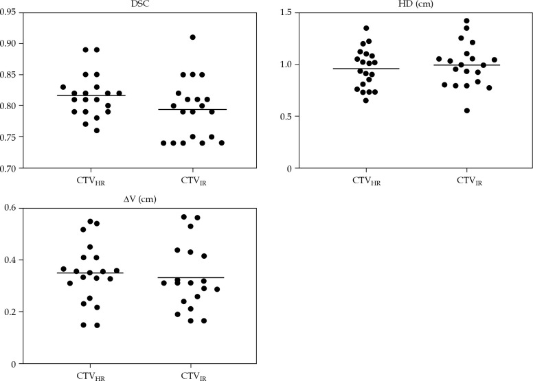
Average age of 20 patients included was 57.8 years. Width, thickness, and volumes of CT-based CTV (CTV-CT) were significantly overestimated compared with those of MRI-based CTV (CTV-MR). Mean values of dice similarity coefficient (DSC), Hausdorff distance (HD), and centroid distance (ΔV) of CTV were 0.82 cm, 0.96 cm, and 0.35 cm, respectively. Dose values of CTV-CT were significantly lower compared with those of CTV-MR. Concerning OARs, geometrical and dosimetric values on CT were comparable to those on MRI.

CONCLUSIONS

The delineated ranges of CTV were significantly over-estimated on CT compared with MRI. D and D of CTV-CT were lower than CTV-MR. DSC and ΔV of CTV and CTV were similar to each other; however, there was a difference in terms of HD. CT images regarding pre-BT MR images for delineating were not enough and MRI fusion is still required.

摘要

目的

本研究的目的是比较和评估基于计算机断层扫描(CT)的靶区勾画与基于磁共振成像(MRI)的宫颈癌高剂量率近距离放疗(HDR-BT)的准确性。

材料与方法

收集并评估20例局部晚期宫颈癌患者的数据。将基于MRI的高危临床靶区(CTV)的尺寸、适形性和剂量参数以及危及器官(OARs)的D、D和D与基于CT的进行比较。

结果

纳入的20例患者的平均年龄为57.8岁。与基于MRI的CTV(CTV-MR)相比,基于CT的CTV(CTV-CT)的宽度、厚度和体积被显著高估。CTV的骰子相似系数(DSC)、豪斯多夫距离(HD)和质心距离(ΔV)的平均值分别为0.82 cm、0.96 cm和0.35 cm。CTV-CT的剂量值显著低于CTV-MR。关于OARs,CT上的几何和剂量学值与MRI上的相当。

结论

与MRI相比,CT上CTV的勾画范围被显著高估。CTV-CT的D和D低于CTV-MR。CTV和CTV的DSC和ΔV彼此相似;然而,在HD方面存在差异。用于勾画的BT前MR图像的CT图像不够,仍需要MRI融合。

https://cdn.ncbi.nlm.nih.gov/pmc/blobs/9e4d/7690230/26bc8b891183/JCB-12-41568-g003.jpg
https://cdn.ncbi.nlm.nih.gov/pmc/blobs/9e4d/7690230/9847610baa7f/JCB-12-41568-g001.jpg
https://cdn.ncbi.nlm.nih.gov/pmc/blobs/9e4d/7690230/aeac11cdf024/JCB-12-41568-g002.jpg
https://cdn.ncbi.nlm.nih.gov/pmc/blobs/9e4d/7690230/26bc8b891183/JCB-12-41568-g003.jpg
https://cdn.ncbi.nlm.nih.gov/pmc/blobs/9e4d/7690230/9847610baa7f/JCB-12-41568-g001.jpg
https://cdn.ncbi.nlm.nih.gov/pmc/blobs/9e4d/7690230/aeac11cdf024/JCB-12-41568-g002.jpg
https://cdn.ncbi.nlm.nih.gov/pmc/blobs/9e4d/7690230/26bc8b891183/JCB-12-41568-g003.jpg

相似文献

1
Comparison of computed tomography- and magnetic resonance imaging-based target delineation for cervical cancer brachytherapy.基于计算机断层扫描和磁共振成像的宫颈癌近距离放疗靶区勾画比较
J Contemp Brachytherapy. 2020 Aug;12(4):367-374. doi: 10.5114/jcb.2020.98117. Epub 2020 Aug 21.
2
Dosimetric impact of target definition in brachytherapy for cervical cancer - Computed tomography and trans rectal ultrasound versus magnetic resonance imaging.宫颈癌近距离治疗中靶区定义的剂量学影响——计算机断层扫描和经直肠超声与磁共振成像的比较
Phys Imaging Radiat Oncol. 2022 Mar 4;21:126-133. doi: 10.1016/j.phro.2022.02.002. eCollection 2022 Jan.
3
Value of Magnetic Resonance Imaging Without or With Applicator in Place for Target Definition in Cervix Cancer Brachytherapy.磁共振成像(MRI)在宫颈癌近距离放疗中置管前后靶区勾画的价值
Int J Radiat Oncol Biol Phys. 2016 Mar 1;94(3):588-97. doi: 10.1016/j.ijrobp.2015.09.023.
4
Comparison of impact of target delineation of computed tomography- and magnetic resonance imaging-guided brachytherapy on dose distribution in cervical cancer.计算机断层扫描和磁共振成像引导下近距离放射治疗的靶区勾画对宫颈癌剂量分布影响的比较
J Contemp Brachytherapy. 2018 Oct;10(5):418-424. doi: 10.5114/jcb.2018.78993. Epub 2018 Oct 15.
5
Hybrid TRUS/CT with optical tracking for target delineation in image-guided adaptive brachytherapy for cervical cancer.基于光学追踪的混合式经直肠超声/计算机断层扫描在宫颈癌图像引导自适应近距离放疗中的靶区勾画。
Strahlenther Onkol. 2020 Nov;196(11):983-992. doi: 10.1007/s00066-020-01656-2. Epub 2020 Jul 3.
6
Comparison of CTV and organs at risk contours between TRUS and MR images in IB cervical cancers: a proof of concept study.在 IB 期宫颈癌中,比较经直肠超声和磁共振图像的 CTV 和危及器官勾画:一项概念验证研究。
Radiat Oncol. 2020 Apr 6;15(1):73. doi: 10.1186/s13014-020-01516-4.
7
Effect of tumor dose, volume and overall treatment time on local control after radiochemotherapy including MRI guided brachytherapy of locally advanced cervical cancer.肿瘤剂量、体积及总治疗时间对局部晚期宫颈癌放化疗(包括MRI引导下近距离放疗)后局部控制的影响。
Radiother Oncol. 2016 Sep;120(3):441-446. doi: 10.1016/j.radonc.2016.05.014. Epub 2016 Jun 24.
8
Diffusion-weighted MRI in image-guided adaptive brachytherapy: Tumor delineation feasibility study and comparison with GEC-ESTRO guidelines.图像引导下适形近距离放射治疗中的扩散加权磁共振成像:肿瘤勾画可行性研究及与GEC-ESTRO指南的比较
Brachytherapy. 2017 Sep-Oct;16(5):956-963. doi: 10.1016/j.brachy.2017.05.010. Epub 2017 Jun 30.
9
Dynamics of High Risk Clinical Target Volume reduction during Brachytherapy and impact on its coverage in patients with inoperable cervical cancer.近距离放疗中高危临床靶区体积变化的动力学及其对不可手术宫颈癌患者靶区覆盖的影响。
Neoplasma. 2018 Mar 14;65(3):425-430. doi: 10.4149/neo_2018_170113N30.
10
Impact of different treatment plans on EQD for intracavitary brachytherapy of cervical cancer.不同治疗计划对宫颈癌腔内近距离放疗等效剂量的影响。
J Egypt Natl Canc Inst. 2021 Oct 4;33(1):28. doi: 10.1186/s43046-021-00084-2.

引用本文的文献

1
Impact of MRI-based planning in transperineal interstitial brachytherapy for vaginal-involving tumors.基于磁共振成像(MRI)的计划在经会阴间质近距离放疗治疗累及阴道肿瘤中的应用
J Contemp Brachytherapy. 2024 Oct;16(5):335-343. doi: 10.5114/jcb.2024.144683. Epub 2024 Oct 30.
2
Fully automated segmentation of clinical target volume in cervical cancer from magnetic resonance imaging with convolutional neural network.基于卷积神经网络的宫颈癌磁共振成像临床靶区全自动分割
J Appl Clin Med Phys. 2022 Sep;23(9):e13725. doi: 10.1002/acm2.13725. Epub 2022 Jul 27.

本文引用的文献

1
Combined Ultrasound and Cone Beam CT Improves Target Segmentation for Image Guided Radiation Therapy in Uterine Cervix Cancer.联合超声和锥形束 CT 可改善宫颈癌图像引导放疗中的靶区勾画。
Int J Radiat Oncol Biol Phys. 2019 Jul 1;104(3):685-693. doi: 10.1016/j.ijrobp.2019.03.003. Epub 2019 Mar 11.
2
Comparison of impact of target delineation of computed tomography- and magnetic resonance imaging-guided brachytherapy on dose distribution in cervical cancer.计算机断层扫描和磁共振成像引导下近距离放射治疗的靶区勾画对宫颈癌剂量分布影响的比较
J Contemp Brachytherapy. 2018 Oct;10(5):418-424. doi: 10.5114/jcb.2018.78993. Epub 2018 Oct 15.
3
Use of a novel atlas for muscles of mastication to reduce inter observer variability in head and neck radiotherapy contouring.
使用新型咀嚼肌图谱减少头颈部放疗勾画中的观察者间变异性。
Radiother Oncol. 2019 Jan;130:56-61. doi: 10.1016/j.radonc.2018.10.030. Epub 2018 Nov 9.
4
A Prospective Comparison of Computed Tomography with Transrectal Ultrasonography Assistance and Magnetic Resonance Imaging-Based Target-Volume Definition During Image Guided Adaptive Brachytherapy for Cervical Cancers.一种前瞻性比较研究:在影像引导自适应近距离放射治疗宫颈癌时,与经直肠超声辅助和基于磁共振成像的靶区定义相比。
Int J Radiat Oncol Biol Phys. 2018 Dec 1;102(5):1448-1456. doi: 10.1016/j.ijrobp.2018.05.080. Epub 2018 Jun 7.
5
Automatic segmentation of the clinical target volume and organs at risk in the planning CT for rectal cancer using deep dilated convolutional neural networks.使用深度扩张卷积神经网络在直肠癌计划 CT 中自动分割临床靶区和危及器官。
Med Phys. 2017 Dec;44(12):6377-6389. doi: 10.1002/mp.12602. Epub 2017 Oct 28.
6
What is the optimal number of library plans in ART for locally advanced cervical cancer?ART 中局部晚期宫颈癌的最佳放疗计划数是多少?
Radiother Oncol. 2017 Dec;125(3):470-477. doi: 10.1016/j.radonc.2017.08.033. Epub 2017 Sep 19.
7
Evaluation of segmentation methods on head and neck CT: Auto-segmentation challenge 2015.头部和颈部 CT 分割方法评估:2015 年自动分割挑战赛。
Med Phys. 2017 May;44(5):2020-2036. doi: 10.1002/mp.12197. Epub 2017 Apr 21.
8
Comparison of computed tomography and magnetic resonance imaging in cervical cancer brachytherapy: A systematic review.宫颈癌近距离放疗中计算机断层扫描与磁共振成像的比较:一项系统评价
Brachytherapy. 2017 Mar-Apr;16(2):353-365. doi: 10.1016/j.brachy.2016.11.001. Epub 2016 Dec 10.
9
Value of Magnetic Resonance Imaging Without or With Applicator in Place for Target Definition in Cervix Cancer Brachytherapy.磁共振成像(MRI)在宫颈癌近距离放疗中置管前后靶区勾画的价值
Int J Radiat Oncol Biol Phys. 2016 Mar 1;94(3):588-97. doi: 10.1016/j.ijrobp.2015.09.023.
10
Hybrid (CT/MRI based) vs. MRI only based image-guided brachytherapy in cervical cancer: Dosimetry comparisons and clinical outcome.宫颈癌中基于混合(CT/MRI)与仅基于MRI的图像引导近距离放射治疗:剂量学比较与临床结果
Brachytherapy. 2016 Jan-Feb;15(1):40-8. doi: 10.1016/j.brachy.2015.09.002. Epub 2015 Oct 23.