Division of Diabetes, Metabolism and Endocrinology, Department of Internal Medicine, Iwate Medical University.
Division of Ultra-high Field MRI, Institute for Biomedical Sciences, Iwate Medical University.
J Atheroscler Thromb. 2019 Dec 1;26(12):1045-1053. doi: 10.5551/jat.48553. Epub 2019 Mar 15.
It remains unclear whether elevated low-density lipoprotein cholesterol (LDL-C) is a risk factor for cerebral vascular disease. Familial hypercholesterolemia (FH) is the most appropriate model for understanding the effects of excess LDL-C because affected individuals have inherently high levels of circulating LDL-C. To clarify the effects of hypercholesterolemia on cerebral small vessel disease (SVD), we investigated cerebrovascular damage in detail due to elevated LDL-C using high resolution brain magnetic resonance imaging (MRI) in patients with FH.
Twenty-eight patients with FH and 35 healthy controls underwent 7T brain MRI. The prevalence of SVD and arterial structural changes were determined in each group.
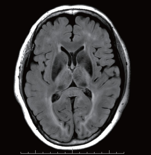
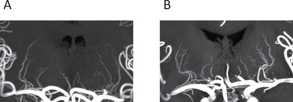
The prevalence of periventricular hyperintensity (PVH) was significantly higher (control, 0% vs. FH, 14.2%, p=0.021) and deep white matter intensity tended to be more frequent in FH patients than in controls. The prevalence of SVD in patients with forms of cerebral damage, such as lacunar infarction, PVH, deep white matter hyperintensities (DWMH), microbleeding, and brain atrophy, was significantly higher among FH patients (control, n=2, 5.7% vs. FH, n=7, 25.0%, p<0.001, chi-square test). The tortuosity of major intracranial arteries and the signal intensity of lenticulostriate arteries were similar in the two groups. In FH patients, as the grade of PVH progressed, several atherosclerosis risk factors, such as body mass index, blood pressure, and triglyceride level, showed ever worsening values.
These results obtained from FH patients revealed that persistently elevated LDL-C leads to cerebral PVH. It is necessary in the management of FH to pay attention not only to the development of coronary heart disease but also to the presence of cerebral SVD.
目前尚不清楚升高的低密度脂蛋白胆固醇(LDL-C)是否是脑血管疾病的危险因素。家族性高胆固醇血症(FH)是理解过量 LDL-C 影响的最合适模型,因为受影响的个体本身具有高水平的循环 LDL-C。为了阐明高胆固醇血症对脑小血管疾病(SVD)的影响,我们使用 7T 脑磁共振成像(MRI)详细研究了 FH 患者因 LDL-C 升高导致的脑血管损伤。
28 例 FH 患者和 35 例健康对照者接受了 7T 脑 MRI 检查。确定每组 SVD 和动脉结构变化的发生率。
脑室周围高信号(PVH)的发生率明显更高(对照组 0% vs. FH 组 14.2%,p=0.021),且 FH 患者的深部白质高信号更常见。伴有腔隙性梗死、PVH、深部白质高信号(DWMH)、微出血和脑萎缩等脑损伤形式的 FH 患者的 SVD 发生率明显高于对照组(对照组 n=2,5.7% vs. FH 组 n=7,25.0%,p<0.001,卡方检验)。两组主要颅内动脉的迂曲度和纹状体动脉信号强度相似。在 FH 患者中,随着 PVH 分级的进展,一些动脉粥样硬化危险因素,如体重指数、血压和甘油三酯水平,显示出越来越差的值。
这些从 FH 患者中获得的结果表明,持续升高的 LDL-C 可导致脑 PVH。在 FH 的管理中,不仅要注意冠心病的发生,还要注意脑 SVD 的存在。