• 文献检索
  • 文档翻译
  • 深度研究
  • 学术资讯
  • Suppr Zotero 插件Zotero 插件
  • 邀请有礼
  • 套餐&价格
  • 历史记录
应用&插件
Suppr Zotero 插件Zotero 插件浏览器插件Mac 客户端Windows 客户端微信小程序
定价
高级版会员购买积分包购买API积分包
服务
文献检索文档翻译深度研究API 文档MCP 服务
关于我们
关于 Suppr公司介绍联系我们用户协议隐私条款
关注我们

Suppr 超能文献

核心技术专利:CN118964589B侵权必究
粤ICP备2023148730 号-1Suppr @ 2026

文献检索

告别复杂PubMed语法,用中文像聊天一样搜索,搜遍4000万医学文献。AI智能推荐,让科研检索更轻松。

立即免费搜索

文件翻译

保留排版,准确专业,支持PDF/Word/PPT等文件格式,支持 12+语言互译。

免费翻译文档

深度研究

AI帮你快速写综述,25分钟生成高质量综述,智能提取关键信息,辅助科研写作。

立即免费体验

微出血与遗忘型轻度认知障碍和早期阿尔茨海默病的皮质下血管周围间隙扩大共存:7T MRI 研究。

Microbleeds colocalize with enlarged juxtacortical perivascular spaces in amnestic mild cognitive impairment and early Alzheimer's disease: A 7 Tesla MRI study.

机构信息

Brain Center Rudolf Magnus, Department of Neurology, University Medical Center Utrecht, the Netherlands.

J. Philip Kistler Stroke Research Center, Department of Neurology, Massachusetts General Hospital and Harvard Medical School, Boston, MA, USA.

出版信息

J Cereb Blood Flow Metab. 2020 Apr;40(4):739-746. doi: 10.1177/0271678X19838087. Epub 2019 Mar 19.

DOI:10.1177/0271678X19838087
PMID:30890076
原文链接:https://pmc.ncbi.nlm.nih.gov/articles/PMC7074594/
Abstract

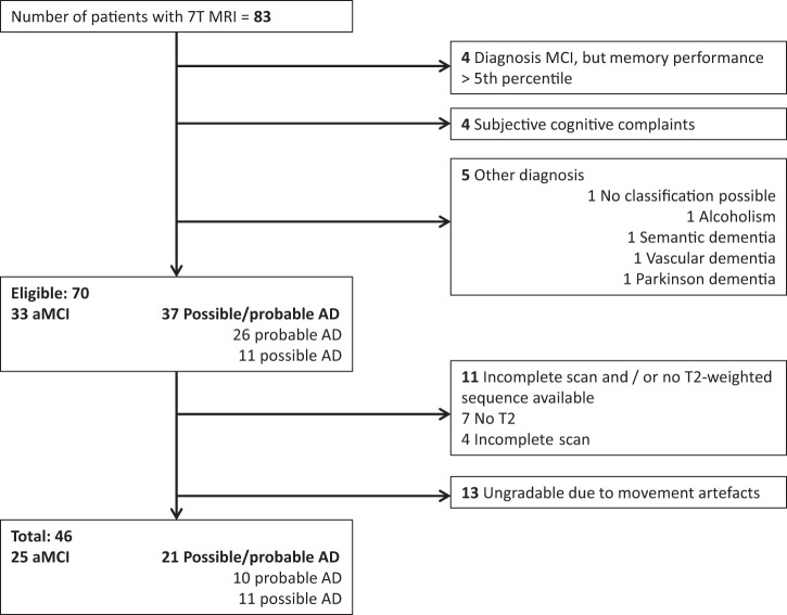
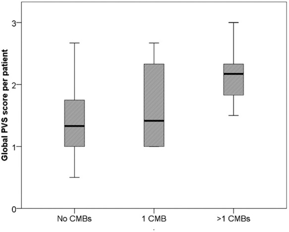
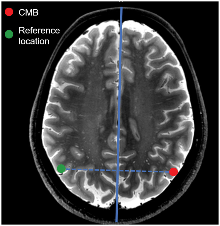
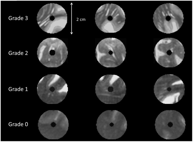
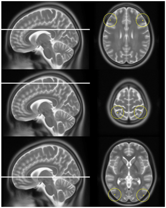
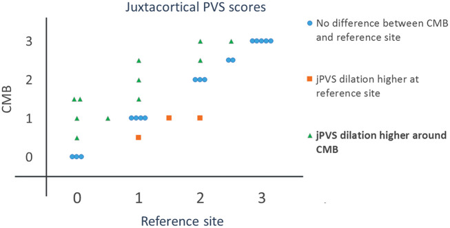
MRI-visible perivascular spaces (PVS) in the semioval centre are associated with cerebral amyloid angiopathy (CAA), but it is unknown if PVS co-localize with MRI markers of CAA. To examine this, we assessed the topographical association between cortical cerebral microbleeds (CMBs) - as an indirect marker of CAA - and dilatation of juxtacortical perivascular spaces (jPVS) in 46 patients with amnestic mild cognitive impairment (aMCI) or early Alzheimer's disease (eAD). The degree of dilatation of jPVS <1 cm around each cortical CMBs was compared with a similar reference site (no CMB) in the contralateral hemisphere, using a 4-point scale. Also, jPVS dilatation was compared between patients with and without cortical CMBs. Eleven patients (24%) had cortical CMBs [total=35, median=1, range=1-14] of whom five had >1 cortical CMBs. The degree of jPVS dilatation was higher around CMBs than at the reference sites [Wilcoxon signed rank test, Z = 2.2,  = 0.03]. Patients with >1 cortical CMBs had a higher degree of jPVS dilation [median=2.2, IQR = 1.8-2.3] than patients without cortical CMBs [median=1.4, IQR = 1.0-1.8],  = 0.02. We found a topographical association between a high degree of jPVS dilatation and cortical CMBs, supporting a common underlying pathophysiology - most likely CAA.

摘要

磁共振成像(MRI)可见的半卵圆中心血管周围间隙(PVS)与脑淀粉样血管病(CAA)有关,但尚不清楚 PVS 是否与 CAA 的 MRI 标志物共存。为了研究这一点,我们评估了皮质脑微出血(CMB)——作为 CAA 的间接标志物——与皮质下血管周围间隙扩张(jPVS)之间的拓扑关联,共纳入 46 例遗忘型轻度认知障碍(aMCI)或早期阿尔茨海默病(eAD)患者。使用 4 分制量表比较每个皮质 CMB 周围 jPVS 的扩张程度与对侧半球相似的参考部位(无 CMB)。此外,比较了有和无皮质 CMB 患者的 jPVS 扩张情况。11 例患者(24%)有皮质 CMB [总数=35,中位数=1,范围=1-14],其中 5 例有>1 个皮质 CMB。与参考部位相比,CMB 周围的 jPVS 扩张程度更高[Wilcoxon 符号秩检验,Z=2.2,=0.03]。有>1 个皮质 CMB 的患者 jPVS 扩张程度更高[中位数=2.2,IQR=1.8-2.3],而无皮质 CMB 的患者 jPVS 扩张程度较低[中位数=1.4,IQR=1.0-1.8],=0.02。我们发现,jPVS 扩张程度较高与皮质 CMB 之间存在拓扑关联,支持潜在的共同病理生理学机制——最有可能是 CAA。

https://cdn.ncbi.nlm.nih.gov/pmc/blobs/b530/7168785/cc08632e9379/10.1177_0271678X19838087-fig6.jpg
https://cdn.ncbi.nlm.nih.gov/pmc/blobs/b530/7168785/518b2a0833c2/10.1177_0271678X19838087-fig1.jpg
https://cdn.ncbi.nlm.nih.gov/pmc/blobs/b530/7168785/6442029ab81e/10.1177_0271678X19838087-fig2.jpg
https://cdn.ncbi.nlm.nih.gov/pmc/blobs/b530/7168785/65eb2566a0a6/10.1177_0271678X19838087-fig3.jpg
https://cdn.ncbi.nlm.nih.gov/pmc/blobs/b530/7168785/062c72972fd6/10.1177_0271678X19838087-fig4.jpg
https://cdn.ncbi.nlm.nih.gov/pmc/blobs/b530/7168785/a9d448664383/10.1177_0271678X19838087-fig5.jpg
https://cdn.ncbi.nlm.nih.gov/pmc/blobs/b530/7168785/cc08632e9379/10.1177_0271678X19838087-fig6.jpg

相似文献

1
Microbleeds colocalize with enlarged juxtacortical perivascular spaces in amnestic mild cognitive impairment and early Alzheimer's disease: A 7 Tesla MRI study.微出血与遗忘型轻度认知障碍和早期阿尔茨海默病的皮质下血管周围间隙扩大共存:7T MRI 研究。
J Cereb Blood Flow Metab. 2020 Apr;40(4):739-746. doi: 10.1177/0271678X19838087. Epub 2019 Mar 19.
2
Comparison of cerebrospinal fluid profiles in Alzheimer's disease with multiple cerebral microbleeds and cerebral amyloid angiopathy-related inflammation.阿尔茨海默病合并多发性脑微出血及脑淀粉样血管病相关炎症患者的脑脊液特征比较
J Neurol. 2017 Feb;264(2):373-381. doi: 10.1007/s00415-016-8362-2. Epub 2016 Dec 20.
3
Hypertensive Arteriopathy and Cerebral Amyloid Angiopathy in Patients with Cognitive Decline and Mixed Cerebral Microbleeds.认知功能下降伴混合性脑微出血患者的高血压性血管病变和脑淀粉样血管病。
J Alzheimers Dis. 2020;78(4):1765-1774. doi: 10.3233/JAD-200992.
4
Cortical microinfarcts in patients with multiple lobar microbleeds on 3 T MRI.3T MRI 上多发性脑叶微出血患者的皮质微梗死。
J Neurol. 2019 Aug;266(8):1887-1896. doi: 10.1007/s00415-019-09350-9. Epub 2019 May 2.
5
Centrum Semiovale Perivascular Space and Amyloid Deposition in Spontaneous Intracerebral Hemorrhage.半卵圆中心血管周围间隙与自发性脑出血中的淀粉样沉积。
Stroke. 2021 Jul;52(7):2356-2362. doi: 10.1161/STROKEAHA.120.032139. Epub 2021 Apr 20.
6
Cerebral small vessel disease in patients with spontaneous cerebellar hemorrhage.自发性小脑出血患者的脑小血管病。
J Neurol. 2019 Mar;266(3):625-630. doi: 10.1007/s00415-018-09177-w. Epub 2019 Jan 8.
7
MRI-visible perivascular spaces in cerebral amyloid angiopathy and hypertensive arteriopathy.脑淀粉样血管病和高血压性动脉病中磁共振成像可见的血管周围间隙
Neurology. 2017 Mar 21;88(12):1157-1164. doi: 10.1212/WNL.0000000000003746. Epub 2017 Feb 22.
8
Cognitive Hemorrhagic Onset in Cerebral Amyloid Angiopathy: Neuroimaging Features.脑淀粉样血管病认知性出血性卒中:神经影像学特征。
Curr Alzheimer Res. 2023;20(4):267-276. doi: 10.2174/1567205020666230713151211.
9
MRI-visible perivascular space location is associated with Alzheimer's disease independently of amyloid burden.MRI 可见的血管周围间隙位置与阿尔茨海默病有关,与淀粉样蛋白负担无关。
Brain. 2017 Apr 1;140(4):1107-1116. doi: 10.1093/brain/awx003.
10
Cerebellar Microbleed Distribution Patterns and Cerebral Amyloid Angiopathy.小脑微出血的分布模式与脑淀粉样血管病。
Stroke. 2019 Jul;50(7):1727-1733. doi: 10.1161/STROKEAHA.119.024843. Epub 2019 Jun 4.

引用本文的文献

1
Altered Clearance in Alzheimer's Disease and Cerebral Amyloid Angiopathy.阿尔茨海默病和脑淀粉样血管病中的清除率改变
Neuroimaging Clin N Am. 2025 May;35(2):277-286. doi: 10.1016/j.nic.2024.12.004. Epub 2025 Feb 19.
2
Sex-Specific Patterns of Cerebral Atrophy and Enlarged Perivascular Spaces in Patients with Cerebral Amyloid Angiopathy and Dementia.性别特异性脑萎缩和脑淀粉样血管病伴痴呆患者的血管周围间隙扩大模式。
AJNR Am J Neuroradiol. 2023 Jul;44(7):792-798. doi: 10.3174/ajnr.A7900. Epub 2023 Jun 8.
3
Role of endocrine PACAP in age-related diseases.

本文引用的文献

1
Cerebrovascular function in presymptomatic and symptomatic individuals with hereditary cerebral amyloid angiopathy: a case-control study.遗传性脑淀粉样血管病无症状和有症状个体的脑血管功能:一项病例对照研究。
Lancet Neurol. 2017 Feb;16(2):115-122. doi: 10.1016/S1474-4422(16)30346-5. Epub 2016 Dec 16.
2
Association of Perivascular Localization of Aquaporin-4 With Cognition and Alzheimer Disease in Aging Brains.血管周围水通道蛋白-4 定位与衰老大脑认知功能和阿尔茨海默病的关系。
JAMA Neurol. 2017 Jan 1;74(1):91-99. doi: 10.1001/jamaneurol.2016.4370.
3
Vascular, glial, and lymphatic immune gateways of the central nervous system.
内分泌 PACAP 在与年龄相关的疾病中的作用。
Front Endocrinol (Lausanne). 2023 Mar 9;14:1118927. doi: 10.3389/fendo.2023.1118927. eCollection 2023.
4
Role of Vitamin D Deficiency in the Pathogenesis of Cardiovascular and Cerebrovascular Diseases.维生素 D 缺乏在心血管和脑血管疾病发病机制中的作用。
Nutrients. 2023 Jan 9;15(2):334. doi: 10.3390/nu15020334.
5
Arteriolar neuropathology in cerebral microvascular disease.脑微血管病的小动脉神经病理学。
Neuropathol Appl Neurobiol. 2023 Feb;49(1):e12875. doi: 10.1111/nan.12875.
6
Effect of cerebral small vessel disease on cognitive impairment in Parkinson's disease.脑小血管病对帕金森病认知障碍的影响。
Acta Neurol Belg. 2023 Apr;123(2):487-495. doi: 10.1007/s13760-022-02078-w. Epub 2022 Sep 12.
7
Neuroimaging at 7 Tesla: a pictorial narrative review.7特斯拉的神经成像:图文并茂的综述
Quant Imaging Med Surg. 2022 Jun;12(6):3406-3435. doi: 10.21037/qims-21-969.
8
Imaging perivascular space structure and function using brain MRI.利用脑 MRI 成像研究血管周围空间的结构和功能。
Neuroimage. 2022 Aug 15;257:119329. doi: 10.1016/j.neuroimage.2022.119329. Epub 2022 May 21.
9
Association of Apolipoprotein E ɛ4 Allele with Enlarged Perivascular Spaces.载脂蛋白 E ɛ4 等位基因与血管周围空间扩大的关联。
Ann Neurol. 2022 Jul;92(1):23-31. doi: 10.1002/ana.26364. Epub 2022 Apr 27.
10
Cognitive Impairment of Patient With Neurological Cerebrovascular Disease Using the Artificial Intelligence Technology Guided by MRI.基于MRI引导的人工智能技术对神经脑血管疾病患者认知功能障碍的研究
Front Public Health. 2022 Mar 3;9:813641. doi: 10.3389/fpubh.2021.813641. eCollection 2021.
中枢神经系统的血管、神经胶质和淋巴免疫通道。
Acta Neuropathol. 2016 Sep;132(3):317-38. doi: 10.1007/s00401-016-1606-5. Epub 2016 Aug 13.
4
Clinical features of high-degree centrum semiovale-perivascular spaces in cerebral amyloid angiopathy.脑淀粉样血管病中高度半卵圆中心周围血管间隙的临床特征
J Neurol Sci. 2016 Aug 15;367:89-94. doi: 10.1016/j.jns.2016.05.040. Epub 2016 May 20.
5
Sporadic Cerebral Amyloid Angiopathy: Pathophysiology, Neuroimaging Features, and Clinical Implications.散发性脑淀粉样血管病:病理生理学、神经影像学特征及临床意义。
Semin Neurol. 2016 Jun;36(3):233-43. doi: 10.1055/s-0036-1581993. Epub 2016 May 23.
6
Perivascular spaces on 7 Tesla brain MRI are related to markers of small vessel disease but not to age or cardiovascular risk factors.7特斯拉脑部磁共振成像上的血管周围间隙与小血管疾病标志物相关,但与年龄或心血管危险因素无关。
J Cereb Blood Flow Metab. 2016 Oct;36(10):1708-1717. doi: 10.1177/0271678X16648970. Epub 2016 May 6.
7
Cerebral amyloid angiopathy severity is linked to dilation of juxtacortical perivascular spaces.脑淀粉样血管病的严重程度与皮质旁血管周围间隙的扩张有关。
J Cereb Blood Flow Metab. 2016 Mar;36(3):576-80. doi: 10.1177/0271678X15620434. Epub 2015 Dec 7.
8
Prevalence of Brain Microbleeds in Alzheimer Disease: A Systematic Review and Meta-Analysis on the Influence of Neuroimaging Techniques.阿尔茨海默病中脑微出血的患病率:关于神经影像学技术影响的系统评价和荟萃分析
AJNR Am J Neuroradiol. 2016 Feb;37(2):215-22. doi: 10.3174/ajnr.A4525. Epub 2015 Dec 3.
9
Assessment of blood flow velocity and pulsatility in cerebral perforating arteries with 7-T quantitative flow MRI.使用7-T定量血流磁共振成像评估脑穿支动脉的血流速度和搏动性。
NMR Biomed. 2016 Sep;29(9):1295-304. doi: 10.1002/nbm.3306. Epub 2015 Apr 27.
10
White matter perivascular spaces on magnetic resonance imaging: marker of cerebrovascular amyloid burden?磁共振成像上的脑白质血管周围间隙:脑血管淀粉样蛋白负荷的标志物?
Stroke. 2015 Jun;46(6):1707-9. doi: 10.1161/STROKEAHA.115.009090. Epub 2015 Apr 23.