Adamo Ana M, Liu Xiuzhen, Mathieu Patricia, Nuttall Johnathan R, Supasai Suangsuda, Oteiza Patricia I
Department of Biological Chemistry and IQUIFIB (UBA-CONICET), Facultad de Farmacia y Bioquimica, Universidad de Buenos Aires, Buenos Aires, Argentina.
Department of Nutrition and Department of Environmental Toxicology, University of California, Davis, Davis, CA, United States.
Front Cell Neurosci. 2019 Mar 1;13:62. doi: 10.3389/fncel.2019.00062. eCollection 2019.
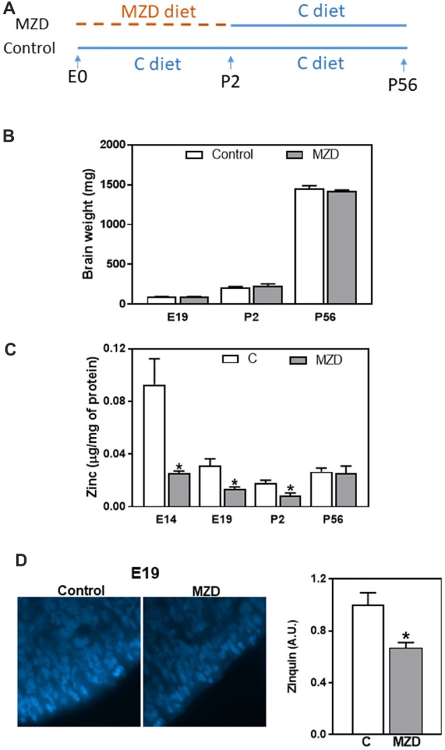
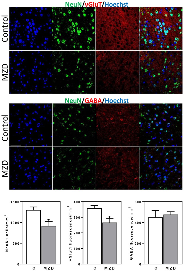
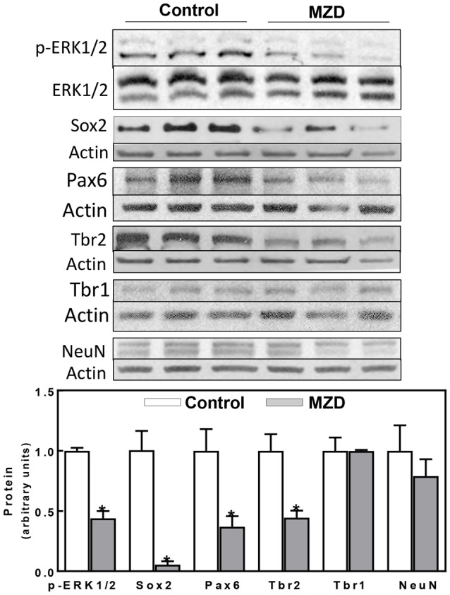
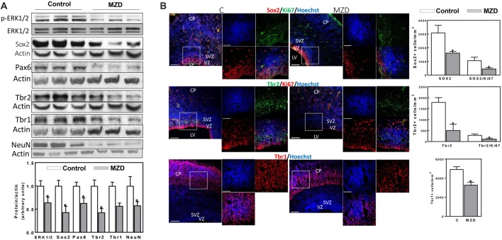
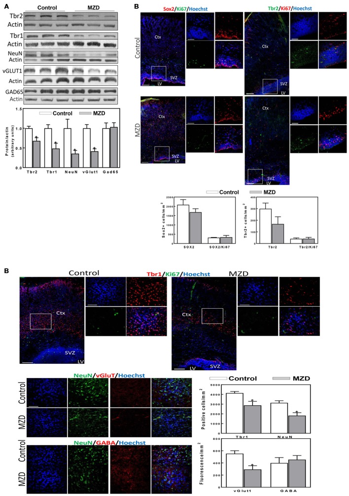
During pregnancy, a decreased availability of zinc to the fetus can disrupt the development of the central nervous system leading to defects ranging from severe malformations to subtle neurological and cognitive effects. We previously found that marginal zinc deficiency down-regulates the extracellular signal-regulated kinase 1/2 (ERK1/2) signaling pathway and affects neural progenitor cell (NPC) proliferation. This study investigated if marginal zinc deficiency during gestation in rats could disrupt fetal neurogenesis and affect the number and specification of neurons in the adult offspring brain cortex. Rats were fed a marginal zinc deficient or adequate diet throughout gestation and until postnatal day (P) 2, and subsequently the zinc adequate diet until P56. Neurogenesis was evaluated in the offspring at embryonic day (E)14, E19, P2, and P56 measuring parameters of NPC proliferation and differentiation by Western blot and/or immunofluorescence. At E14 and E19, major signals (i.e., ERK1/2, Sox2, and Pax6) that stimulate NPC proliferation and self-renewal were markedly downregulated in the marginal zinc deficient fetal brain. These alterations were associated to a lower number of Ki67 positive cells in the ventricular (VZs) and subventricular zones (SVZs). Following the progression of NPCs into intermediate progenitor cells (IPCs) and into neurons, Pax6, Tbr2 and Tbr1 were affected in the corresponding areas of the brain at E19 and P2. The above signaling alterations led to a lower density of neurons and a selective decrease of glutamatergic neurons in the young adult brain cortex exposed to maternal marginal zinc deficiency from E14 to P2. Current results supports the concept that marginal zinc deficiency during fetal development can disrupt neurogenesis and alter cortical structure potentially leading to irreversible neurobehavioral impairments later in life.
在孕期,胎儿可利用的锌减少会干扰中枢神经系统的发育,导致从严重畸形到细微神经和认知影响等一系列缺陷。我们之前发现,边缘性锌缺乏会下调细胞外信号调节激酶1/2(ERK1/2)信号通路,并影响神经祖细胞(NPC)的增殖。本研究调查了大鼠孕期边缘性锌缺乏是否会扰乱胎儿神经发生,并影响成年后代大脑皮层中神经元的数量和分化。大鼠在整个孕期及出生后第2天(P2)饲喂边缘性锌缺乏或充足的饮食,随后直至出生后第56天(P56)饲喂锌充足的饮食。在胚胎期第14天(E14)、E19、P2和P56对后代的神经发生进行评估,通过蛋白质免疫印迹法和/或免疫荧光法测量NPC增殖和分化的参数。在E14和E19时,边缘性锌缺乏的胎儿大脑中刺激NPC增殖和自我更新的主要信号(即ERK1/2、Sox2和Pax6)明显下调。这些改变与脑室(VZs)和脑室下区(SVZs)中Ki67阳性细胞数量减少有关。随着NPCs向中间祖细胞(IPCs)和神经元的转变,E19和P2时大脑相应区域的Pax6、Tbr2和Tbr1受到影响。上述信号改变导致在E14至P2暴露于母体边缘性锌缺乏的年轻成年大脑皮层中神经元密度降低,且谷氨酸能神经元选择性减少。目前的结果支持这样的观点,即胎儿发育期间的边缘性锌缺乏会扰乱神经发生并改变皮层结构,可能导致后期生活中不可逆转的神经行为障碍。